ISMRM Honors 2022

Fellow of the Society - ISMRM

Shreyas S. Vasanawala, M.D., Ph.D.

For his contributions as both a clinician and a scientist which span the development of hardware, image reconstruction/processing, to its implementation in the clinics, specifically in the areas of compressed sensing in pediatric body MRI, pediatric coil development and motion correction.

Junior Fellows

Congyu Liao and Nan Wang named Junior Fellows 

Summa Cum Laude

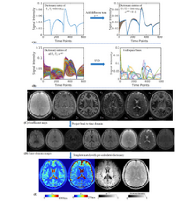
3D Diffusion-Prepared MRF (3DM) With
Cardiac Gating For Rapid High Resolution Whole-
Brain T1, T2, Proton Density And Diffusivity Mapping

Xiaozhi Cao, Congyu Liao, Zheng Zhong, Erpeng Dai, Siddharth Srinivasan Iyer, Ariel J Hannum, Mahmut Yurt, Stefan Skare, and Kawin Setsompop

In this work, a diffusion preparation was implemented in a 3D spiral-projection MRF framework to introduce additional diffusion weighting. Using MRF dictionary with diffusion terms, it enables whole-brain T1, T2, PD and additional diffusivity mapping with 1.25-mm isotropic resolution within 3min. To improve the image quality, cardiac gating and low-resolution navigator were also implemented to mitigate the signal variation caused by motion during diffusion encoding. Subspace reconstruction was used along with LLR regularization to improve the reconstruction conditioning as well as SNR.


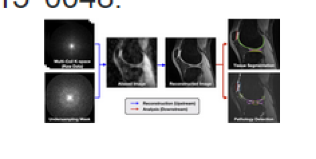
SKM-TEA: A Dataset For Accelerated MRI
Reconstruction With Dense Image Labels For Quantitative
Clinical Evaluation

Arjun D Desai, Andrew M Schmidt, Elka B Rubi2, Christopher M Sandino, Marianne S Black, Valentina Mazzoli, Kathryn J Stevens, Robert Boutin, Christopher Ré, Garry E Gold, Brian A Hargreaves, and Akshay S Chaudhari

While deep-learning-based MRI reconstruction and image analysis methods have shown promise, few have been translated to clinical practice. This may be a result of (1) paucity of end-to-end datasets that enable comprehensive evaluation from reconstruction to analysis and (2) discordance between conventional validation metrics and clinically useful endpoints. Here, we present the Stanford Knee MRI with Multi-Task Evaluation (SKM-TEA), a dataset of 155 clinical quantitative 3D knee MRI scans with k-space data, DICOM images, and dense tissue segmentation and pathology annotations to facilitate clinically relevant, comprehensive benchmarking of the MRI workflow. Dataset, code, and trained baselines are available at https://github.com/StanfordMIMI/skm-tea.


Smaller MRgFUS Lesions That Overlap Patient-
Fit Normative VIM—Precentral Tracts Improve Quality-Of-
Life Outcomes In Essential Tremor

Yosef Chodakiewitz1, David Arnold Purger1, Alan Rehn Wang1, Daniel Barbosa1, Lior Lev Tov1, Anjali Datta1, Rachelle Bitton1, Jennifer McNab1, Vivek Buch1, and Pejman Ghanouni1

While Focused-Ultrasound thalamotomy has proven effective at reducing tremor, traditional targeting methods can be suboptimal at balancing primary tremor-reduction outcomes against undesired side-effects. The traditional “canonical” technique involves an indirect method which applies a non-individualized stereotactic coordinate atlas towards identifying the presumed approximate location of VIM thalamus, the ablation target; the canonical lesion is empirically grown in size based-on dynamic intraoperative feedback from an awake patient, until the surgeon judges that an appropriate balance of tremor-reduction and side-effect risk has been achieved. We propose optimized methods to define and monitor the ideal anatomical ablation for optimized tremor-reduction/Quality-of-Life balancing.


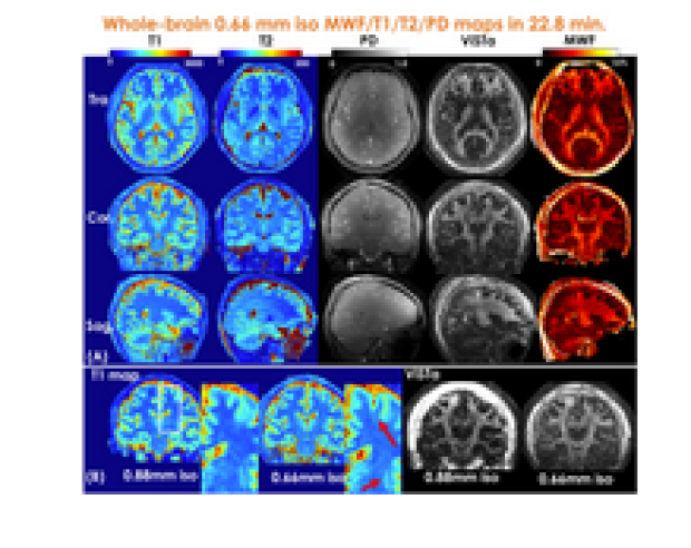
Mesoscale Myelin-Water Fraction And T1/T2
/PD Mapping Through Optimized 3D ViSTa-MRF And
Stochastic Reconstruction With Preconditioning

Congyu Liao, Xiaozhi Cao, Siddharth Srinivasan Iyer, Zihan Zhou, Yunsong Liu, Justin Haldar, Mahmut Yurt, Ting Gong, Zhe Wu, Hongjian He, Jianhui Zhong, Adam Kerr, and Kawin Setsompop

In this work, we developed ViSTa-MRF, which combined Visualization of Short Transverse relaxation time component (ViSTa) technique with MR Fingerprinting (MRF), to achieve high-fidelity whole-brain myelin-water fraction (MWF) and T1/T2/PD mapping at sub-millimeter isotropic resolution on a clinical 3T scanner. To achieve fast acquisition and memory-efficient reconstruction, the ViSTa-MRF sequence leverages an optimized 3D tiny-golden-angle-shuffling (TGAS) spiral-projection acquisition and stochastic subspace reconstruction with optimized k-space diagonal preconditioning. With the proposed ViSTa-MRF method, high-fidelity direct MWF mapping was achieved without a need for multi-compartment fitting.


Magna Cum Laude

Designing A Clinical Decision Support System For
MRI Radiology Titles Using Machine Learning Techniques And
Electronic Medical Records

Peyman Shokrollahi, Juan M. Zambrano Chaves, Jonathan P.H. Lam, Avishkar Sharma, Debashish Pal, Naeim Bahrami, Akshay S. Chaudhari, and Andreas M. Loening

The use of inappropriate radiology protocols introduces risk of missed and incomplete diagnoses, thus endangering patient health, potentially prolonging treatment, and increasing healthcare costs. A clinical decision support system based on machine learning and electronic medical records of patients undergoing MRI was developed to predict radiology titles and their probabilities for radiologist review. A cumulative F1-score of ~85% was obtained for the top three predicted titles. The proposed system can guide physicians toward selecting appropriate titles and alert radiologists of potentially inappropriate selections, thereby improving imaging utility and increasing diagnostic accuracy, which favors better patient outcomes. 


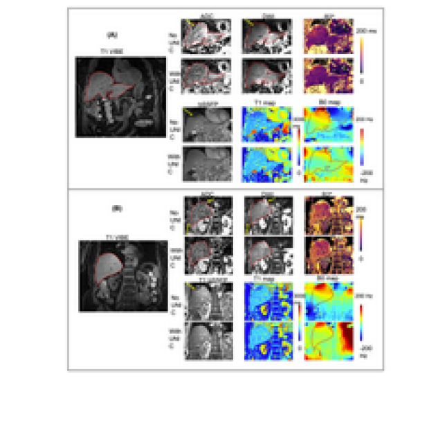
Integrated High-Order B0 Shimming For
Multiparametric Quantitative Liver Imaging at 3T Using A UNIfied Coil (UNIC)

Nan Wang, Fardad M. Serry, Michael Ocasio, Yibin Xie, Yuheng Huang, Xinqi Li, Pei Han, Tianle Cao, Sen Ma, Fei Han, Matthew Minton, Yubin Cai, Yujie Shan, Xiaoming Bi, Anthony G. Christodoulou, Hsin-Jung Yang, Debiao Li, and Hui Han

Liver MRI shows promise for rich morphologic and physiologic information. B0 inhomogeneity causes off-resonance spread at the liver-lung interfaces, degrading image quality and quantification accuracy for DWI, T1, and T2*/R2*. A novel high-order shim coil (UNIC) was built, minimizing the decoupling between the two overlapped shim and RF arrays, allowing the free design of shim loop topology in proximity of the target organ. Improved shimming from UNIC is demonstrated with increased FOV in liver imaging, showing significantly reduced distortion in liver DWI, increased image quality score of MOLLI T1 map, and reduced B0-offset-induced ADC, R2*, and B0 standard deviations.

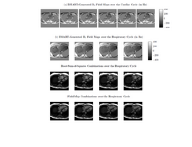
Field-Map Combination Method for Phase-Cycled
bSSFP using Inherent B0 Mapping

Anjali Datta, Dwight Nishimura, and Corey Baron

Phase-cycled bSSFP enables off-resonance-robust bSSFP imaging. Conventional phase-cycle combination methods weight brighter phase-cycles more, assuming that passband signal is brighter than off-resonant signal. However, near-band signal can be hyperintense, most notably for flowing spins, so traditional methods fail to mitigate these artifacts. Incorporating knowledge of Bo enables inclusion of only passband signal, and exclusion of dark bands and near-band hyperintense artifacts. Using a golden-angle radial trajectory for a free-breathing, phase-cycled acquisition enables reconstruction of a Bo-map time series without any additional scans. This facilitates field-map-based combination throughout the respiratory and cardiac cycles, resulting in substantially reduced hyperintense artifacts than root-sum-of-squares.