Research Areas
Cancer and autoimmune diseases are among the most common causes of disability and death worldwide. Addressing this medical challenge to help our patients requires understanding of disease pathogenesis and progression to enable its early detection. Our lab takes advantage of optical, chemical, and biological properties of diseased tissue to further our understanding of disease pathogenesis and progression while we contribute to developing molecular imaging technologies for early cancer detection and theragnostics.
Fluorescence and Raman Augmented Endoscopy

Our current project emphasizes transitioning near-infrared human endoscopes and tumor-targeting molecular agent into clinical workflows. We are developing fluorescence-ready and Raman-ready flexible endoscopes to target (pre)cancerous tissue in patients; our approach can detect cell numbers that are an order of magnitude smaller than existing imaging standards (CT, MRI, PET). Using this methodology, we enable precise disease detection via guided biopsy sampling to monitor disease progression to achieve more accurate post-treatment cancer surveillance.
Funding: Innovative Medicines Accelerator (IMA) at Stanford University
Collaborators: Matthew Bogyo, Andrew Shelton, George Poultsides, George Fisher, Erqi Pollum (Stanford University, USA), Vergent, Curadel
Vibrational Spectroscopy for Theragnostic Approaches in the Gastrointestinal Tract
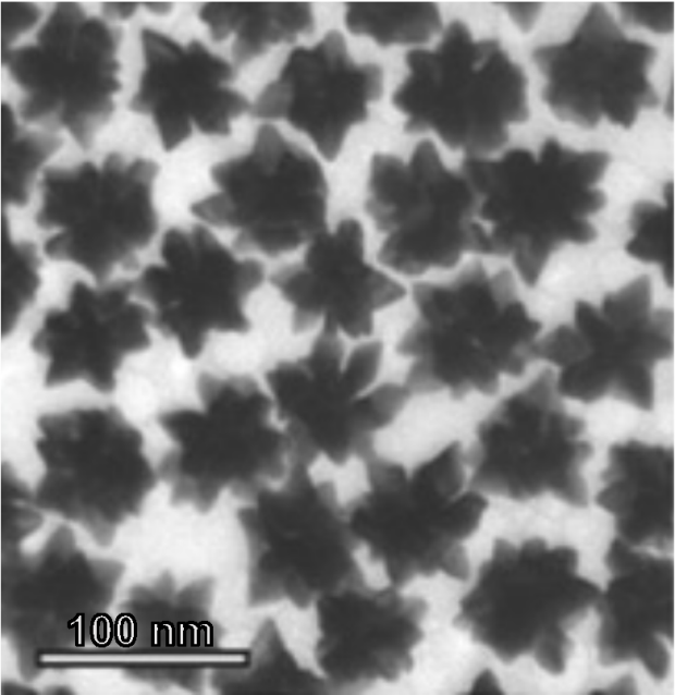
From the range of vibrational spectroscopies available, we investigate using Raman spectroscopy as a laser-mediated imaging modality to measure spectral fingerprints that emerge from vibrational active modes present in the tissue’s chemical composition. Using surface enhanced resonance Raman scattering (SERRS) nanoparticles and nanostars binding to cancerous and dysplastic tissue, we can not only reliably detect pre-cancerous lesions in the gastrointestinal tract but also induce photothermal ablation of the either dysplastic or malignant tissue that is associated with cancer pathogenesis and progression. In summary, our nanostar technology combines diagnostic and therapeutic functionality into a single theragnostic agent.
Our lab also investigates the feasibility of infrared spectroscopy to optimize sample selection for other imaging and *omics technologies.
Funding: National Institute of Health, National Cancer Institute, Maternal and Child Health Research Institute (MCHRI) at Stanford University, Canary Center for Early Detection
Collaborators: Alexander Vahrmeijer, Stefan Harmsen (Leiden University, Netherlands); Garry Nolan (Stanford University, USA)
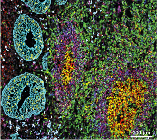
Exploring Cellular Neighborhoods and Tissue Architectures in Gastrointestinal Diseases