MIPS Collaborative Research Develops PET Tracer to Image Bacterial Infection
May 26, 2020
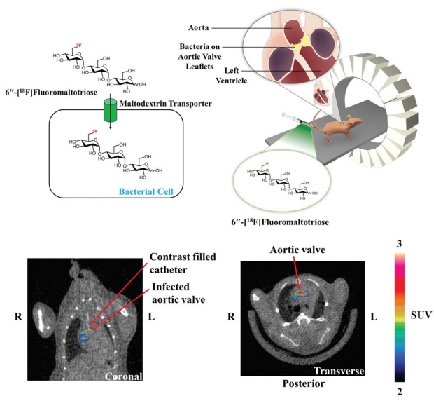
In this study, researchers developed a positron emission tomography-computed tomography (PET-CT) based strategy for directly imaging bacteria in an infective endocarditis (IE) mouse model using the novel probe 6''-[18F]Fluoromaltotriose. This probe targets the maltodextrin transporter, a carrier system that is exclusive to bacteria and not expressed in mammalian cells. To our knowledge, this is the first time that a fluorine-18 PET tracer has been used to specifically image bacterial infection of the heart valves with high sensitivity and specificity in an animal model. Responses to antibiotic therapy could also be assessed using 6′′-[18F]Fluoromaltotriose PET-CT. Plans are currently underway to translate the 6''-[18F]Fluoromaltotriose tracer into the clinic for imaging patients with IE and cardiovascular device infections.
This project was a multi-disciplinary collaboration between scientists, engineers, and physicians at Stanford University, the Institute for Quantitative Health Sciences and Engineering at Michigan State University, Verily Life Sciences and Google Health.