Whole-body cell tracking using CellGPS
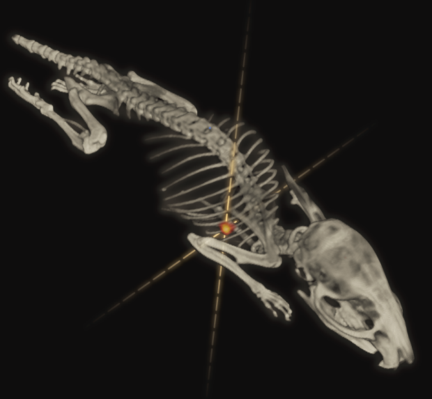
We are building new methods for tracking moving cells anywhere in the body of living sujects. The technique, which is called CellGPS, uses positron emission tomography (PET) to non-invasively measure the 3D position of individual cells as a function of time, and is being applied to study how cells navigate the body's circulatory system. The general methodology can be applied to understand the efficacy of cell-based therapies, including cancer immunotherapy and regenerative medicine, or to study biological processes involving cell migration, such as developmental biology or cancer metastasis. This project blends disciplines such as imaging physics, algorithm development, radiochemistry, and biomedical applications.
Related Publications
Jung KO, Kim TJ, Yu JH, Rhee S, Zhao W, Byunghang Ha, Red-Horse K, Gambhir SS & Pratx G, "Whole-body tracking of single cells via positron emission tomography", Nat. Biomed. Eng., 2020
Ouyang Y, Kim TJ & Pratx G, "Evaluation of a BGO-Based PET System for Single-Cell Tracking Performance by Simulation and Phantom Studies," Mol. Imaging 15, pp. 1-8, 2016
Lee KS, Kim TJ, Pratx G, “Single-Cell Tracking with PET using a Novel Trajectory Reconstruction Algorithm,” IEEE Trans Med Imag, pp. 994-1003, 2014
Microphysiological models for nuclear medicine
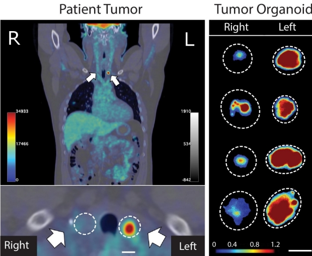
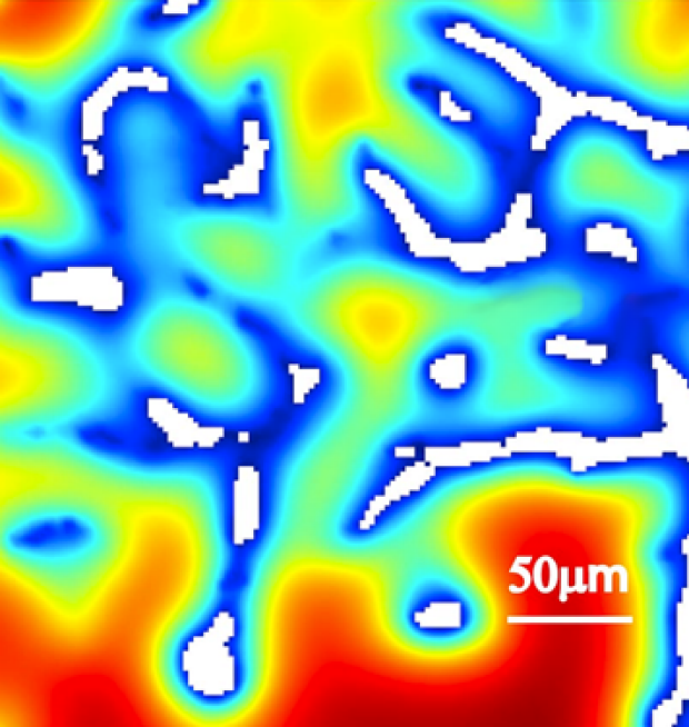
Microphysiological tumor models (μPTMs) are miniature tumors derived from patient tissues that recapitulate the essential hallmarks of solid tumors. We are developping novel approaches to probe these models using clinical radiotracers at an unprecedented level of spatial resolution. This work is based on the development of radioluminescence microscopy, a method that was pioneered here at Stanford and that can image radionuclide-labeled molecules in a standard microscopy environment, down to the level of single cells. Using this new workflow, we are able to incorporate translational imaging endpoints in pre-clinical organoid studies, which brides the gap between in vitro research and clinical trials.
Related publications
Khan S, Shin JH, Ferri V, Cheng N, Noel JE, Kuo C, Sunwoo JB & Pratx G, "High-resolution positron emission microscopy of patient-derived tumor organoids," Nat. Communications 12, pp. 5883
Khan S, Kim S, Yang YP & Pratx G, "High-resolution radioluminescence microscopy of FDG uptake in an engineered 3D tumor-stoma model," Eur. J. Nuc. Med. Mol. Imaging 48(11), 3400-3407
Türkcan S, Kiru L, Naczynski D, Sasportas LS & Pratx G, "Lactic acid accumulation in the tumor microenvironment suppresses 18F- FDG uptake," Cancer Res. 79(2), pp. 410-419, 2019
Kiru L, Kim TK, Shen B, Chin FT & Pratx G, "Single-cell imaging using radioluminescence microscopy reveals unexpected binding target for [18F]HFB," Mol. Imaging Biol. 20(3), pp. 378-387, 2017
Kim TJ, Türkcan S & Pratx G, “Modular low-light microscope for imaging cellular bioluminescence and radioluminescence”, Nat. Protoc. 12, pp. 1055–1076, 2017
Sengupta D & Pratx G, "Single-cell characterization of FLT uptake with radioluminescence microscopy," J. Nucl. Med. 57(7), pp. 1136-1140, 2016
Natarajan A, Türkcan S, Gambhir SS & Pratx G, "A multiscale framework for imaging radiolabeled therapeutics," Mol. Pharm. 2015 12 (12), pp. 4554–4560, 2015
Sengupta D, Miller S, Marton Z, Chin F, Nagarkar V, & Pratx G. "Bright Lu2O3:Eu Thin-Film Scintillators for High-Resolution Radioluminescence Microscopy.", Adv Healthc Mater 4(14), pp. 2064-2070, 2015
Pratx G, Chen K, Sun C, Axente M, Sasportas L, Carpenter C & Xing L, "High-Resolution Radioluminescence Microscopy of FDG Uptake by Reconstructing the Beta Ionization Track", J. Nucl. Imag. 54(10) pp.1841-1846, 2013
Pratx G, Chen K, Sun C, Martin L, Carpenter CM, Olcott PD & Xing L, "Radioluminescence microscopy: Measuring the heterogeneous uptake of radiotracers in single living cells", PLOS One7(10), e46285, 2012
FLASH radiotherapy
Radiation therapy is highly effective in many different cancers. However, the adverse effects of the treatment on healthy organs near the tumor remain a concern. In this context, “FLASH” radiation, which delivers the treatment in the blink of an eye, is being investigated for its ability to spare normal tissues with no concomitant decrease in tumor control. The Physical Oncology Lab participates in ongoing efforts at Stanford to develop, apply and investigate FLASH radiotherapy. Specifically, we are interested in elucidating the role of oxygen as a potential sparing mechanism. In these studies, both theoretical and experimental models are used to tease out the relative contribution of different factors to radiation responses. These studies provide a greater mechanistic understanding of the FLASH effect critical for clinical translation in humans.
Related Publications
Ha B, Liang K, Liu C, Melemenidis S, Manjappa R, Viswanathan V, Das N, Ashraf R, Lau B, Soto S, Graves EE, Rao J, Loo BW, and Pratx G, “Real-time optical oximetry during FLASH radiotherapy using a phosphorescent nanoprobe,” Radiother. Oncol. (in press)
Cui S & Pratx G, “3D computational model of oxygen depletion kinetics in brain vasculature during FLASH RT and its implications for in vivo oximetry experiments,” Med. Phys. 49(6), pp. 3914-3925
Khan S, Bassenne M, Wang J, Manjappa R, Melemenidis S, Breitkreutz DY, Maxim PG, Xing L, Loo BW, & Pratx G, “Multicellular spheroids as in vitro models of oxygen depletion during FLASH irradiation,” Int. J. Radiat. Oncol. Biol. Phys. 110(3), pp. 833-844
Pratx G & Kapp DS, “A computational model of radiolytic oxygen depletion during FLASH irradiation and its effect on the oxygen enhancement ratio,” Phys. Med. Biol. 64, pp. 185005, 2019
Pratx G & Kapp DS, "Ultra-high dose rate FLASH irradiation may spare hypoxic stem cell niches in normal tissues," Int. J. Radiat. Oncol. Biol. Phys. 105(1), pp. 190-192, 2019
Microfluidics assays for oncology
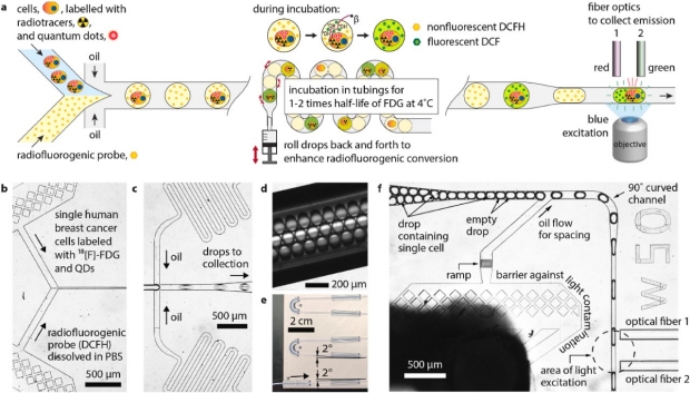
Our research uses microfluidics technology precisely manipulate small volumes of reagents and cells for “lab-on-a-chip” assays. We have developped a number of different systems, including a single-cell radiotracer uptake assay (known as flow radiocytometry), a cell uptake kinetic assay, and a novel approach based on mechanoporation to label cells with a radiolabel for in vivo tracking studies.
Related Publications
Nejadnik H, Jung KO, Theruvath AJ, Kiru L, Liu A, Wu W, Sulchek T, Pratx G & Daldrup-Link HE, "Instant labeling of therapeutic cells for multimodality imaging," Theranostics 10, 6024-6034
Jung KO, Theruvath AJ, Nejadnik H, Liu A, Xing L, Sulchek T, Daldrup-Link HE & Pratx G, “Mechanoporation enables rapid and efficient radiolabeling of stem cells for PET imaging,” Sci. Rep. 12, pp. 2955
Ha B, Kim TJ, Moon EJ, Giaccia AJ & Pratx G, "Flow Radiocytometry Using Droplet Optofluidics," Biosens. Bioelectron. 194, pp. 113565
Kim TJ, Ha B, Bick AD, Kim M, Tang SKY & Pratx G, “Microfluidics-Coupled Radioluminescence Microscopy for In Vitro Radiotracer Kinetic Studies," Anal. Chem. 93 (10), pp. 4425-4433
Sengupta D, Mongersun A, von Eyben R, Abbyad P & Pratx G, “Multiplexed measurements of single-cell FDG uptake and lactate release using droplet microfluidics,”Technol. Cancer. Res. Treat., pp. 1-9
Gallina ME, Kim TJ, Shelor M, Vasquez J, Mongersun A, Kim M, Tang SKY, Abbyad P & Pratx G, “Towards a droplet-based single-cell radiometric assay”, Anal. Chem. 89 (12), pp 6472–6481, May 2017