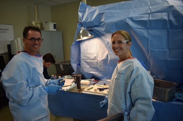
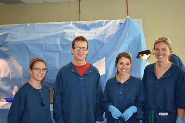
Sports Medicine Research
At Stanford, we understand that athletics are a vital part of life for adolescents. The development of physical motor skills, and the teamwork and communication tools acquired through sports and physical activity are important for well rounded adolescent growth and development. Through expert patient care- from your child’s clinical visits and surgical experience, to the physical therapy and return to play preparation- our goal is to provide your child with the best tools for a successful and timely return to their athletic endeavors. Our pediatric sports research team studies the best surgical treatments, care pathways, and rehabilitation programs to get your child back to 100% in the most efficient way possible.


Publications
-
Age, Sex, and BMI Differences Related to Repairable Meniscal Tears in Pediatric and Adolescent Patients.
The American journal of sports medicine
2023: 3635465221145939
More
Abstract
The incidence of meniscus tears and ACL tears in pediatric patients continues to rise, bringing to question the risk factors associated with these injuries. As meniscus tears are commonly repaired in pediatric populations, the epidemiology of repairable meniscus tears is an important for consideration for surgeons evaluating treatment options.To describe meniscal tear patterns in pediatric and adolescent patients who underwent meniscal repair across multiple institutions and surgeons, as well as to evaluate the relationship between age, sex, and body mass index (BMI) and their effect on the prevalence, type, and displacement of repaired pediatric meniscal tears.Case series; Level of evidence, 4.Data within a prospective multicenter cohort registry for quality improvement, Sport Cohort Outcome Registry (SCORE), were reviewed to describe repaired meniscal tear patterns. All consecutive arthroscopic meniscal repairs from participating surgeons in patients aged <19 years were analyzed. Tear pattern, location, and displacement were evaluated by patient age, sex, and BMI. A subanalysis was also performed to investigate whether meniscal tear patterns differed between those occurring in isolation or those occurring with a concomitant anterior cruciate ligament (ACL) injury. Analysis of variance was used to generate a multivariate analysis of specified variables. Sex, age, and BMI results were compared across the cohort.There were 1185 total meniscal repairs evaluated in as many patients, which included 656 (55.4%) male and 529 (44.6%) female patients. Patients underwent surgery at a mean age of 15.3 years (range, 5-19 years), with a mean BMI of 24.9 (range, 12.3-46.42). Of the 1185 patients, 816 (68.9%) had ACL + meniscal repair and 369 (31.1%) had isolated meniscal repair. The male patients underwent more lateral tear repairs than the female patients (54.3% to 40.9%; P < .001) and had a lower incidence of medial tear repair (32.1% vs 41.4%; P < .001). Patients with repaired lateral tears had a mean age of 15.0 years, compared with a mean age of 15.4 years for patients with repaired medial or bilateral tears (P = .001). Higher BMI was associated with "complex" and "radial" tear repairs of the lateral meniscus (P < .001) but was variable with regard to medial tear repairs.In pediatric and adolescent populations, the data suggest that the surgical team treating knees with potential meniscal injury should be prepared to encounter more complex meniscal tears, commonly indicated in those with higher BMI, while higher rates of lateral meniscal tears were seen in male and younger patients. Future studies should analyze correlates for meniscal repair survival and outcomes in this pediatric cohort undergoing knee surgery.
View details for DOI 10.1177/03635465221145939
View details for PubMedID 36629442
-
Longitudinal 3T MRI T2 * Mapping of Juvenile Osteochondritis Dissecans (JOCD) Lesions Differentiates Operative from Non-operative Patients - Pilot Study.
Journal of orthopaedic research : official publication of the Orthopaedic Research Society
2022
More
Abstract
Juvenile osteochondritis dissecans (JOCD) is an orthopedic joint disorder of children and adolescents that can lead to premature osteoarthritis. Thirteen patients (mean age: 12.3 years, 4 females), 15 JOCD-affected and five contralateral healthy knees, that had a baseline and a follow-up MRI (mean interval of 8.9 months) and were treated non-operatively during this interval were included. Retrospectively, patients were assigned to operative or non-operative groups based on their electronic medical records. Volumetric mean T2 * values were calculated within regions of interest (progeny lesion, interface, parent bone) and region matched control bone in healthy contralateral knees and condyles. The normalized percentage difference of T2 * between baseline and follow up MRI in non-operative patients significantly increased in progeny lesion (-47.8%, p < 0.001), parent bone (-13.9%, p < 0.001), and interface (-32.3%, p = 0.011), whereas the differences in operative patients were non-significant and below 11%. In non-operative patients, the progeny lesion (p < 0.001) and interface T2 * values (p = 0.012) were significantly higher than control bone T2 * at baseline, but not at follow-up (p = 0.219, p=1.000, respectively). In operative patients, the progeny lesion and interface T2 * values remained significantly elevated compared to the control bone both at baseline (p < 0.001, p < 0.001) and follow-up (p < 0.001, p < 0.001), respectively. Clinical Significance: Longitudinal T2 * mapping differentiated non-healing from healing JOCD lesions following initial non-operative treatment, which may assist in prognosis and improve the ability of surgeons to make recommendations regarding operative versus non-operative treatment. This article is protected by copyright. All rights reserved.
View details for DOI 10.1002/jor.25343
View details for PubMedID 35430743
-
Youth athletes sleep more, practice less, and may lose interest in playing sports due to social distancing mandates.
Preventive medicine reports
2022; 26: 101722
More
Abstract
In-person sport participation was suspended across the United States in the spring of 2020 to slow the spread of the novel coronavirus (COVID-19). The purpose of this study was to survey the impact of COVID-19 on young athletes during a period of social and organized sports restrictions. An anonymous cross-sectional survey study was conducted of youth athletes in the midst of social distancing mandates and consisted of six components: demographics, sport participation, changes in sport-related goals/aspirations, sleep habits, and measures of anxiety and depression. 711 individuals who accessed the survey link yielded 575 (81%) participants with responses available for analysis. All respondents (aged 13.0years) played organized sports, 62% were single-sport athletes, and 74% considered high-level. Participants were training 3.3h less per week, spending more time outside, and 86% of participants continued to train while social distancing. Sleep duration increased (1.2h/night) and sleep quality improved in 29% of young athletes. Additionally, 22% and 28% reported PROMIS anxiety and depression scores characterized as 'mild', 'moderate', or 'severe'. Older single-sport participants reported higher depression scores, while higher anxiety scores were seen in female participants with fewer years played. 10% of young athletes and 20% of teenagers changed their sports-related goals. Training style modifications, decreased training, and increased sleep quantity and quality were positive effects of COVID-19 restrictions, while athletic aspirational changes were undesirable effects. Single-sport athletes may be at greater risk for psychological symptoms when their routine is altered.
View details for DOI 10.1016/j.pmedr.2022.101722
View details for PubMedID 35132371
-
Increased Vascularity in the Neonatal versus Adult Meniscus: Evaluation with Magnetic Resonance Imaging.
Cartilage
2020: 1947603520923143
More
Abstract
Objective. Quantification of meniscus vascularity has been limited with previous techniques, and minimal data exist describing differential vascular zones in the skeletally immature meniscus. The objective of this study is to use quantitative contrast-enhanced magnetic resonance imaging (MRI) to compare meniscal vascularity in neonatal specimens with adults. We hypothesized that the developing meniscus has greater and more uniform vascularity throughout all zones. Design. Ten fresh-frozen human cadaveric knees (5 neonatal, age 0-6 months; 5 adult, 34-67 years) underwent gadolinium-enhanced MRI using an established vascularity quantification protocol. Regions of interest corresponding to peripheral and central zones of the meniscus were identified on pre-contrast coronal images, and signal enhancement within the same regions (normalized against background tissue) was compared between pre- and post-contrast images. Results. The medial and lateral menisci had similar distribution of perfusion (45.8% ± 8.1% medial vs. 54.2% ± 8.1% lateral in neonatal knees; 50.6% ± 11.3% medial vs. 49.4% ± 11.3% lateral in adult knees, P = 0.47). Increased perfusion was demonstrated in the periphery compared with the central zone (2.3:1 in neonatal knees and 3.25:1 in adult knees, P = 0.31). Neonatal specimens demonstrated 6.0-fold greater overall post-contrast meniscal signal enhancement compared with adults (P < 0.0001), with the 0-month specimen demonstrating the greatest proportional signal enhancement. Conclusions. While blood flow to the periphery is greater than to central zones in all menisci, younger menisci receive proportionally greater overall blood flow compared to adults, including to the central zone, suggesting that the immature meniscus is a more biologically active tissue than its adult counterpart.
View details for DOI 10.1177/1947603520923143
View details for PubMedID 32447965
-
Three-Dimensional Quantitative Magnetic Resonance Imaging of Epiphyseal Cartilage Vascularity Using Vessel Image Features: New Insights into Juvenile Osteochondritis Dissecans.
JB & JS open access
2019; 4 (4)
More
Abstract
We introduce a quantitative measure of epiphyseal cartilage vascularity and examine vessel networks during human skeletal maturation. Understanding early morphological changes in the distal femoral condyle is expected to provide information on the pathogenesis of developmental diseases such as juvenile osteochondritis dissecans.Methods: Twenty-two cadaveric knees from donors ranging from 1 month to 10 years of age were included in the study. Images of bone, cartilage, and vascularity were acquired simultaneously with a 3-dimensional gradient-recalled-echo magnetic resonance imaging (MRI) sequence. The secondary ossification center volume and total epiphysis cartilage volume ratio and articular-epiphyseal cartilage complex and epiphyseal cartilage widths were measured. Epiphyseal cartilage vascularity was visualized for 9 data sets with quantitative susceptibility mapping and vessel filtering, resulting in 3-dimensional data to inform vessel network segmentation and to calculate vascular density.Results: Three distinct, non-anastomosing vascular networks (2 peripheral and 1 central) supply the distal femoral epiphyseal cartilage. The central network begins regression as early as 3 months and is absent by 4 years. From 1 month to 3 years, the ratio of central to peripheral vascular area density decreased from 1.0 to 0.5, and the ratio of central to peripheral vascular skeletal density decreased from 0.9 to 0.6. A narrow, peripheral vascular rim was present at 8 years but had disappeared by 10 years. The secondary ossification center progressively acquires the shape of the articular-epiphyseal cartilage complex by 8 years of age, and the central areas of the medial and lateral femoral condyles are the last to ossify.Conclusions: Using cadaveric pediatric knees, we provide quantitative, 3-dimensional measures of epiphyseal cartilage vascular regression during skeletal development using vessel image features. Central areas with both early vascular regression and delayed ossification correspond to predilection sites of juvenile osteochondritis dissecans in this limited case series. Our findings highlight specific vascular vulnerabilities that may lead to improved understanding of the pathogenesis and better-informed clinical management decisions in developmental skeletal diseases.Clinical Relevance: This paradigm shift in understanding of juvenile osteochondritis dissecans etiology and disease progression may critically impact future patient management. Our findings highlight specific vascular vulnerabilities during skeletal maturation in a group of active young patients seen primarily by orthopaedic surgeons and sports medicine professionals.
View details for DOI 10.2106/JBJS.OA.19.00031
View details for PubMedID 32043049
-
The Position of the Popliteal Artery and Peroneal Nerve Relative to the Menisci in Children: A Cadaveric Study.
Orthopaedic journal of sports medicine
2019; 7 (6): 2325967119842843
More
Abstract
Meniscal injury in skeletally immature patients is increasingly reported. During meniscal repair, all-inside devices may protrude beyond the posterior limits of the meniscus, putting the neurovascular structures at risk.The purposes of this study were (1) to examine the relationship between the popliteal artery and the posterolateral and posteromedial aspects of the menisci, (2) to examine the relationship of the peroneal nerve to the posterolateral meniscus, and (3) to develop recommendations for avoiding neurovascular injury during posterior meniscal repair in pediatric patients.Descriptive laboratory study.A total of 26 skeletally immature knee cadaveric specimens (7 females and 19 males) were included. Specimens were divided into age groups: 2-4, 5-8, and 9-11 years. The most posterior extent of the lateral and medial menisci was identified via sagittal and axial views on computed tomography (CT) scans. The shortest distance from the most posterior aspect of the lateral and medial menisci to the popliteal artery and the shortest distance from the posterior aspect of the lateral menisci to the anterior rim of the peroneal nerve were measured, and 3-dimensional models of representative specimens were re-created through use of CT scans.For the age groups 2-4, 5-8, and 9-11 years, the mean minimum distance from the posterolateral meniscus to the popliteal artery was 5.2, 6.7, and 8.2 mm, respectively, and the mean minimum distance from the posteromedial meniscus to the popliteal artery was 12.7, 15.4, and 20.3 mm, respectively. In all groups, the distance between the posteromedial meniscus and the popliteal artery was greater than that between the posterolateral meniscus and the popliteal artery. The mean distance from the peroneal nerve to the lateral meniscus was 13.3, 15.0, and 17.9 mm for the respective groups.Many all-inside meniscal repair devices have sharp tips that penetrate posterior to the meniscus and capsule. This study demonstrated that the distance between the posterior meniscus and popliteal artery is relatively small in pediatric patients, especially for the lateral meniscus region.Because of the higher potential for meniscal healing, meniscal repair is more likely to be performed in pediatric patients. The data in this study regarding the proximity of the lateral meniscus and neurovascular structures may be used to guide safe surgical repair of posterior meniscal tears during the use of all-inside meniscal repair devices in these patients.
View details for DOI 10.1177/2325967119842843
View details for PubMedID 31286001
View details for PubMedCentralID PMC6600506
-
Quadriceps Tendon Graft Anatomy in the Skeletally Immature Patient.
Orthopaedic journal of sports medicine
2019; 7 (7): 2325967119856578
More
Abstract
The quadriceps tendon (QT) is increasingly considered for primary and revision anterior cruciate ligament reconstruction in skeletally immature patients, as it may be harvested as a purely soft tissue graft with considerable tissue volume. Because of distinct rectus tendon (RT) separation from the QT complex, the potential for RT retraction exists and could lead to QT weakness after QT graft harvest.To describe the anatomy of the pediatric QT and clarify decussation of the RT and QT to avoid the risk of delayed RT retraction and QT weakness after QT graft harvest.Descriptive epidemiology study.Nine cadaveric knee specimens (aged 4-11 years) underwent gross dissection. Coronal-plane width and depth of the QT were measured at intervals proximal to the superior pole of the patella at distances of 0.0, 0.5, 1.0, and 1.5 times the length of the patella. The distance was measured from the superior patellar pole to the point of RT separation from the remainder of the deeper/posterior QT.The median patellar length was 28 mm (interquartile range, 26-37 mm). The coronal-plane width of the QT was larger superficially/anteriorly when closest to the patella but wider when measured deeper/posteriorly as the tendon extended proximally. The median distance between the superior pole of the patella and RT separation from the QT was 0.95 times the patellar length. The distance to widening of the deeper/posterior aspect of the QT was 1.14 times the patellar length proximal to the patella.The RT begins a distinct separation from the QT above the superior pole of the patella at a median of 0.95 times the patellar length in skeletally immature specimens. The deeper/posterior aspect of the QT begins to increase in coronal-plane width proximally after a distance of 1.14 times the patellar length above the knee, while the superficial/anterior aspect of the tendon continues to narrow. Awareness of the separation of the RT from the QT, and the coronal-plane width variation aspects of the QT proximally, is important for surgeons utilizing the QT as a graft to avoid inadvertent release of the RT from the rest of the QT complex.
View details for DOI 10.1177/2325967119856578
View details for PubMedID 31321249
View details for PubMedCentralID PMC6624918
MCHRI Poster Presentations - October 2021
Faculty Contact
Dr Kevin Shea

Research Contact
Amin Alayleh
aalayleh@stanford.edu
