Knowles Lab Research
White matter structural change in epilepsy
We are studying how white matter structure changes in relation to seizures in different forms of epilepsy, and how aberrant white matter structure, in turn, shapes neuronal network function. In mouse models, we assess various measures of network synchrony along with longitudinal endpoints including seizure burden, behavior and histological measures of white matter structure.
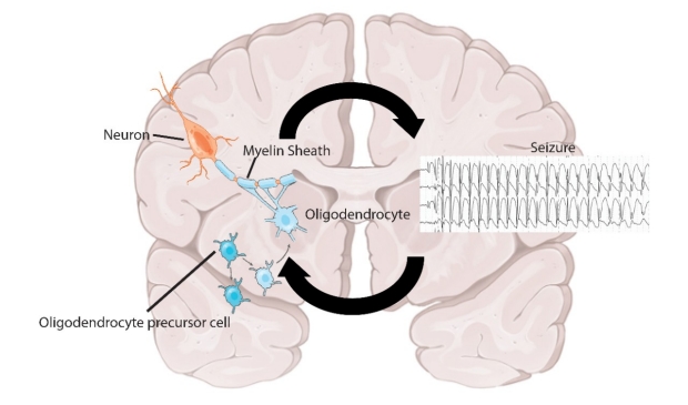
Seizures may induce maladaptive changes in white matter structure and function that, in turn, contribute to epilepsy pathogenesis.
Cellular and molecular mechanisms of maladaptive myelination
What are the molecular mechanisms underlying maladaptive white matter plasticity, and how can they be targeted? We use a range of techniques including in vitro models, optogenetics and transcriptomics to study this question. We are testing novel therapeutic strategies targeting mechanisms of maladaptive myelination in mouse models. We also study how microglia and astrocytes influence pediatric epilepsy.

Representative photomicrographs of oligodendrocyte progenitor cells (left), mature oligodendrocytes (middle). Representative transmission electron micrograph of mid-sagittal callosal axons (right).
Magnetic Resonance Imaging of Epileptogenesis
In collaboration with the lab of Dr. Jennifer McNab at Stanford, we use innovative MR imaging techniques to obtain detailed information related to structural change in brain networks affected by seizures. We use rodent models and plan to expand these studies to children with epilepsy.
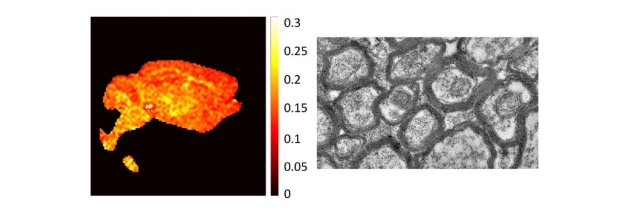
MRI-derived myelin volume fraction map of a mouse brain (left). In vivo measurements of white matter structure made with MRI are validated with electron microscopy (right).
Thalamocortical and white matter dynamics in neonatal and childhood epilepsy
We found that neonates with various genetic forms of epilepsy display convergent white matter abnormalities (Sandoval Karamian et al). In collaboration with Dr. Fiona Baumer and Dr. Chris Lee-Messer, we study how thalamocortical network structure (assessed with imaging) and function (assessed with EEG) change in different forms of childhood epilepsy.
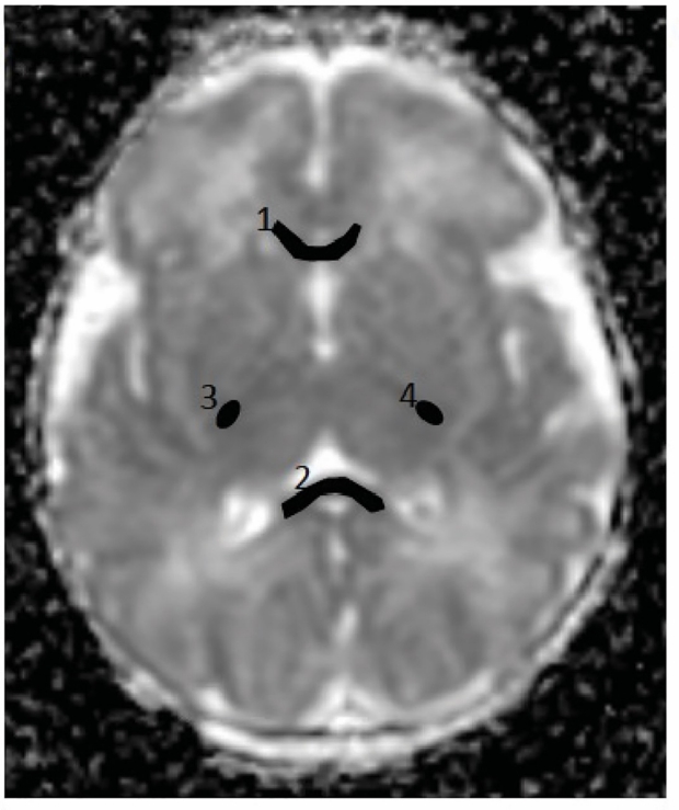
ADC map of neonatal human brain with corpus callosum (1,2) and posterior limbs of internal capsules (3,4) highlighted.