Annual Report 2020
• Advancing optic disc drusen research
> From mother to patient
• Cross-department team effort
• Vision restoration in glaucoma
• Pursuing excellence through diversity, equity, and inclusion
• Advancing clinical research in the age of COVID-19
• Improving ophthalmologic care through artificial intelligence
• Solving corneal blindness with implantable video technology
From mother to patient
A patient’s rare diagnosis plays a pivotal role in a multi-center study
A year ago, Carolyn Miller noticed some sensitivity in her left eye, and her eyelid felt swollen. One week later, she began to experience tingling on the left side of her face, a headache, and pain when she moved her eyes from left to right. She went to Stanford Hospital Emergency Department, where she had tests run, but ultimately, they ruled her initial symptoms as a migraine.
“A week later, my vision in my left eye became cloudy,” Miller said. “It was as if I was looking through grease on a window shield and I couldn’t put the images I was seeing together properly.”
With worsening vision, Miller went back to her local eye specialist. Her doctor suspected the vision problems were being caused by complications in her optic nerve. Being the mother of neuro-ophthalmologist, Shannon Beres, MD, clinical assistant professor of neurology and neurological sciences and ophthalmology at Stanford, Miller asked to be referred to the Byers Eye Institute at Stanford.

Carolyn Miller (left) with her daughter, Shannon Beres, MD, clinical assistant professor of neurology and neurological sciences and ophthalmology.
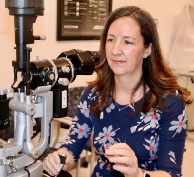
Heather Moss, MD, PhD, diagnosed and treated Carolyn Miller’s rare disease.
A newly discovered disease
Miller came into Byers Eye Institute urgently that day to be seen by her daughter’s neuro-ophthalmology colleague, Heather Moss, MD, PhD, associate professor of ophthalmology and of neurology.
After examining Miller’s vision, Moss ordered an orbital MRI, which revealed inflammation of the optic nerve. Moss diagnosed Miller with optic neuritis, but was still unsure what was causing it.
Moss sent Miller back to the emergency department, where she had a spinal tap and blood work to rule out lymphoma and other inflammatory and infectious diseases. Miller stayed in the hospital and was given a three-day treatment of steroids to decrease her inflammation. After that treatment, Miller’s vision began to return, but as psychedelic patterns and colors.
“I was at my next appointment when the technician asked me to describe what I was seeing,” Miller said. “Out of my left eye it appeared as if she had tattoos with spinning yellow, shooting stars on her arms, but when I covered my left eye those images disappeared.”
After the testing came back and was combined with their experienced serial examinations, Moss and Beres found that the cause of Miller’s optic neuritis was due to an autoimmune neurologic disease called myelin oligodendrocyte glycoprotein (MOG) antibody disease.
“The vivid color hallucinations she experienced in her left eye resulted from the brain trying to compensate for the vision loss,” Moss said. “MOG is a rare disease that was only discovered in the last few years, so her diagnosis is acute in that there is still much to learn about how to tailor treatment for these patients.”
The timing of Miller’s diagnosis was quite serendipitous. She was experiencing symptoms at the same time Moss and Beres were delivering lectures at the Bay Area Ophthalmology Course at Stanford. Their Neuro-Ophthalmology lectures included a focus on optic nerve disorders, and were complemented by invited speakers brought in from across the U.S.
“Dr. Beres and I were doing research on MOG and had invited our collaborator, Dr. John Chen at the Mayo Clinic, where MOG was discovered,” Moss said.
John Chen, MD, PhD, a neuro-ophthalmologist in the Mayo Clinic Department of Ophthalmology, had initiated a multi-center study now in progress at Stanford. Together, leading investigators from around the country are looking at patterns of the disease, the symptoms and imaging features associated with MOG, and studying treatments to prevent relapse of the disease after it is initially treated with steroids.
“I feel fortunate to be a patient where I am at the cutting edge of what is going on for my specific disease.
“I feel fortunate to be a patient where I am at the cutting edge of what is going on for my specific disease,” Miller said. “Dr. Moss and her colleagues have been wonderful. I now have all my doctors here at Stanford, which allows a cohesive and coordinated support of my health profiles. I never feel like something will slip through the cracks.”
After treatment with steroids, Miller’s vision was restored, and she now goes in for monitoring every six months. She has resumed activities that were put on pause when she first lost her vision, like going on walks, as previously her eyes were light-sensitive.
For Beres, this situation was unique for her to observe not just as a physician, but as the patient’s daughter.
“At first it was scary thinking about the short-term and long-term consequences of what this disease could be, but I felt reassured having my mom treated by my colleagues who I know are exceptional,” Beres said.
Miller said she has chosen to keep an optimistic view on the situation and her future. Having lived in the Bay Area her entire life and as a teacher of almost 30 years, now retired, she has developed strong relationships with those in her community, and feels blessed to have a supportive family that all live close to her. She finds it fortuitous to have a daughter who is a neuro-ophthalmologist locally and she is especially grateful for the exceptional care by Moss, who is actively advancing the research for her rare disease through collaborative research with national and international colleagues.
“I have much to be thankful for in my life,” Miller said. “To anyone walking a similar road, I would tell them that keeping a positive attitude and trusting in your support system makes for the best outcomes.”
By KATHRYN SILL
Kathryn Sill is a web and communications specialist for the Byers Eye Institute in the Department of Ophthalmology, at Stanford University School of Medicine. Email her at ksill@stanford.edu.