Overview
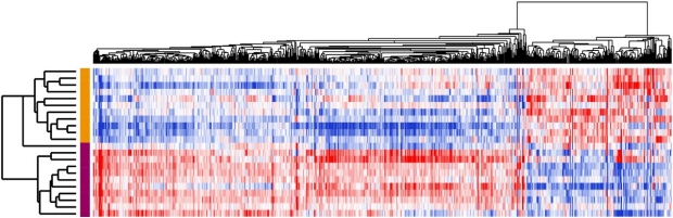
The causes of benign prostatic hyperplasia (BPH) are unknown. Based on our previous work, BPH can be separated into different molecular subtypes based on which genes are expressed. We are now focused on understanding whether these subtypes of BPH are due to different populations of cells in the tissues, specifically the fibroblasts and immune cells. Critical questions we are addressing include: 1) Are there specific fibroblasts responsible for the genesis and growth of BPH? 2) How do immune cells interact and signal with other cells in BPH to contribute to prostate growth? 3) Can these pathways be used in the clinic to guide treatments for BPH or suggest new pathways to prevent or treat BPH? We are investigating these questions through 2 integrated projects and our Biospecimen/Bioimaging core.
Biospecimen/Bioimaging Core
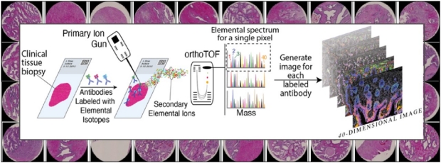
The Biospecimen/Bioimaging Core leverages infrastructure of the Stanford Urology and Pathology Departments to provide services to benefit O’Brien Center investigators in the CAIRIBU Network. The Core procures and provides biospecimens, including fresh, freshly-frozen, and formalin-fixed paraffin-embedded (FFPE) prostate issues. the Core provides tissue characterization services, including histology, tissue microarrays, laser microdissection, and multiplex immunohistochemistry (IHC) using MIBI-TOF (Multiplexed Ion Beam Imaging by Time of Flight). MIBI-TOF is a powerful and robust technology that harnesses metal-tagged antibodies for the simultaneous quantification of 40 or more antibody targets, with superior spatial resolution and dynamic range. The Core also stores and provides portal access to images including prostate MRIs, whole-mount histology and IHC, and MIBI-TOF data, as well as an integrated multiscale data platform.
Research Projects

Project 1
Stromal fibroblasts in BPH pathogenesis
Fibroblasts are a key cell type within the prostate stroma – the lattice of cells and proteins that support the growth and function of the secretory epithelial cells. Fibroblasts play key roles in the development and maintenance of the prostate gland, and are increasingly thought to contribute to the pathogenic enlargement of the prostate in BPH. In recent studies, we found that prostate fibroblasts are not a single cell type, but rather comprise a heterogeneous population of cells with distinct gene expression patterns and presumed functions. The overarching goals of Project 1 are to define the fibroblast cell types in BPH (versus normal prostate), and to determine the key interactions between BPH fibroblasts and epithelium that drive prostate enlargement. To achieve these goals, we will leverage state-of-the art genomics and imaging technologies developed by our groups at Stanford. Findings should inform the origins of BPH, and may suggest new molecular targets for improved therapies.
Project 2
Role of the Immune Microenvironments in BPH
Prostate stroma – the lattice of cells and proteins underlying the secretory epithelial cells – is rich in immune cells. The immune cells are thought to modulate normal tissue maintenance and remodeling, but may also contribute to the abnormal prostate enlargement that characterizes BPH. Supporting that possibility, our recent studies of BPH have uncovered massive overexpression of CXCL13, an immune cell attractant. Leveraging advanced genomic and imaging technologies, the broad goals of Project 2 are to define the immune cell populations in BPH, to understand their roles with respect to prostate enlargement, and to discover the origins of the pathogenic immune response. Findings may suggest new opportunities to manipulate immune cell microenvironment to manage or treat BPH.