A new technology marries ultrasound and photoacoustic imaging techniques to produce a picture showing the anatomy of the prostate, functional details about the gland and molecular information that can help flag cancerous tissue.
September 20, 2019 - By Hanae Armitage
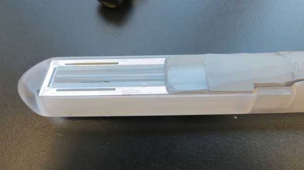
A new device employs ultrasound and photoacoustic imaging techniques to detect prostate cancer.
Courtesy of Sri-Rajasekhar Kothapalli and Sam Gambhir
Getting a close look at the prostate is critical for detecting cancer, but its rather intimate positioning (just in front of the rectum) makes it difficult to image.
Now, Sanjiv “Sam” Gambhir, MD, PhD, professor and chair of radiology, thinks he has a solution: a newly devised hybrid camera.
Traditionally, prostate cancer is detected via prostate cancer-associated blood biomarkers, such as prostate-specific antigen. Doctors often also use ultrasound or MRI to look for physical changes in prostate tissue. Newer techniques that harness positron emission tomography scans can even capture molecular detail, but those tactics are relatively more expensive and use radiation, Gambhir said.
“But the problem is that cancer within the prostate quite often doesn’t lead to any anatomical changes until it’s quite large or has spread beyond the capsule of the prostate into the lymph nodes around it,” he said. “So for decades, we’ve been looking for ways to analyze and image the prostate with greater detail to detect changes earlier on, safely and at relatively low cost.”
Gambhir’s new technology, dubbed the transrectal ultrasound and photoacoustic device, or TRUSPA, marries ultrasound and photoacoustic imaging techniques to simultaneously produce a picture showing the anatomy of the prostate, functional details about the gland and molecular information that can help flag cancerous tissue.
In a proof of principle study, Gambhir and a team of scientists across Stanford, including biologists, engineers and doctors, have demonstrated the value of the instrument in about 20 patients. A paper describing the technology and results was published Aug. 28 in Science Translational Medicine. Gambhir is the senior author of the study, and Sri-Rajasekhar Kothapalli, PhD, is the lead author.
To improve on poke-and-hope
Since ultrasound is already used widely by urologists and generally in human imaging, Gambhir opted to start with that as the foundation of TRUSPA. Typically, if a biomarker such as prostate-specific antigen is elevated in a patient’s blood, doctors then turn to a combination of ultrasound and biopsy, during which they use a needle to take about 20 samples from different regions of the prostate. The technique is rooted in a poke-and-hope theory — as in hopefully you’re sampling the part of the prostate that contains the cancer tissue. But it’s not guaranteed.
TRUSPA takes a different approach, which incorporates an imaging agent that cancer cells readily take up — more so than regular tissue. Then, through photoacoustic molecular imaging (which monitors the absorption of light waves to help characterize tissue type), doctors can see where the cancer cells are located in the prostate. The presence of the imaging agent in tumor tissue changes the way that the light gets absorbed and ultrasound waves are sent back to the device, making it into a sort of flag for cancerous tissue.
“We opted for an imaging agent that was not specific to prostate cancer, but rather to cancerous tissues for our proof of principle,” Gambhir said. That imaging agent was already approved by the Food and Drug Administration, making it an easy starting point. “But the idea moving forward is to heighten precision using a molecularly-targeted photoacoustic molecular imaging agent that binds specifically to prostate cancer cells.”
In the pilot study, the scientists used the device in 20 individuals who had been diagnosed with prostate cancer, looking to see whether it could likewise detect the disease.
“Not only were we able to better understand TRUSPA imaging limitations in the patients, we also were likely seeing tumors that would have otherwise been invisible to conventional prostate ultrasound,” Gambhir said.
In one patient, they were even able to differentiate between malignant and nonmalignant cancer tissue, which was confirmed upon further molecular analysis after the diseased prostate was removed from the patient. Gambhir cautions that this is still a pilot, and that the team needs to test the imaging system much more before concluding that TRUSPA can make these sorts of differentiations broadly. But it is a promising start, he said.
With clear evidence that the concept and technology work in humans, Gambhir and his team are continuing to improve the device, including its spatial resolution and the molecular photoacoustic imaging agents specific for prostate cancer, to enhance the accuracy and sensitivity of tumor detection.
“We’re now starting to explore TRUSPA for detecting ovarian cancer, thyroid cancer and skin cancer too,” he said.
Other Stanford co-authors are Butrus (Pierre) Khuri-Yakub, PhD, professor of electrical engineering; Geoffrey Sonn, MD, assistant professor of urology; Joseph Liao, MD, associate professor of urology; and James Brooks, MD, professor of urology.
-
 Hanae ArmitageHanae Armitage is a science writer in the Office of Communications. Email her at harmitag@stanford.edu.
About Stanford Medicine
Stanford Medicine is an integrated academic health system comprising the Stanford School of Medicine and adult and pediatric health care delivery systems. Together, they harness the full potential of biomedicine through collaborative research, education and clinical care for patients. For more information, please visit med.stanford.edu.

