Fibrous adhesions that form after abdominal surgery may be preventable or treatable, according to Stanford study. Adhesions affect most surgical patients, and treating them costs over $1 billion annually.
November 28, 2018 - By Krista Conger
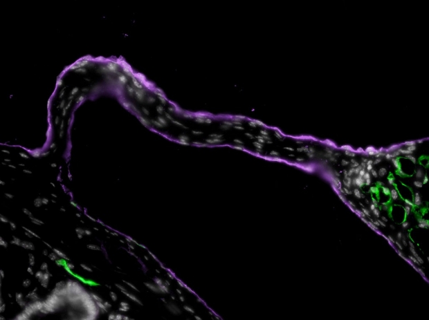
An image of a string adhesion between the liver and the intestine. The cell nuclei are stained white.
Jonathan Tsai
A cellular culprit — as well as a possible treatment — for a common, sometimes life-threating post-surgical complication has been identified by researchers at the Stanford University School of Medicine.
The condition arises when abnormal fibrous connections called adhesions form after abdominal surgery, tethering our normally slippery organs together or anchoring them to the abdominal wall. Symptoms can include chronic pain, female infertility, bowel obstruction and, occasionally, death. According to the National Institutes of Health, the annual cost of treating post-surgical adhesions in the United States surpasses $1 billion.
“This is a very common surgical complication, but it’s not been well-studied,” said Jonathan Tsai, MD, PhD, a former medical student at Stanford and now resident physician at Brigham and Women’s Hospital in Boston. “Until now, it wasn’t even known what cell type was involved in originating the adhesions. Now we’ve come up with a way to isolate the injured tissue before they form the adhesions, and identify the molecular pathways involved.”
The researchers developed and studied a mouse model of adhesion formation to identify the cell responsible for the initial steps. They also showed that an antibody-based therapy could break down those that had already formed. The hope is that similar techniques could help treat post-surgical adhesions in humans.
Tsai is the lead author of the work, which was published Nov. 28 in Science Translational Medicine. Yuval Rinkevich, PhD, a former Stanford postdoctoral scholar, and Irving Weissman, MD, professor of pathology and of developmental biology, share senior authorship of the study. Weissman is the director of the Stanford Institute for Stem Cell Biology and Regenerative Medicine and of Stanford’s Ludwig Cancer Center.
The researchers found that a combination of two antibodies — one that targets the cells responsible for adhesion formation and another that silences a “don’t eat me” signal that cancer cells use to evade the immune system — could significantly reduce the severity of established adhesions in the animals.
“Although we used a mouse model to study adhesion formation,” Weissman said, “we found similar characteristics in adhesions from patients, which makes us think this approach could be translated into the clinic.”
A common complication
Normally the surface of our abdominal organs and the lining of our abdominal cavity are covered with a slippery membrane called mesothelium. The mesothelium allows our organs to glide smoothly past one another when we bend, twist or run. When the mesothelium is disturbed, fibrous connections form between neighboring surfaces, ranging in severity from single threads to vast, immobilizing webs. The NIH estimates that about 93 percent of abdominal surgeries result in adhesions and that about 20 percent of surgical patients will be re-hospitalized for adhesion-related complications.
Irving Weissman
Although the complication is common, it’s not well-understood. Researchers have identified some cell types involved in later steps of the process, but it’s not been known which cell type is responsible for the initial steps. It appears to arise in regions where blood flow is restricted, such as in the tiny pinches of tissue caused by surgical sutures. As a result, less oxygen is delivered to cells in the region — a condition known as hypoxia.
Tsai used a mouse model of the condition to trace the formation of adhesions and the resulting patterns of gene expression in the mesothelium.
“We found that adhesions arise from cells of the mesothelium after injury,” Tsai said. “By tracing the patterns of gene expression, we were able to come up with a cellular ‘family tree’ for these fibrotic tissues and identify the biological pathways involved.”
Tsai and his colleagues found that, in mice, cells of the mesothelium respond to hypoxia by making a protein called HIF1alpha. This in turn promotes the expression of other proteins essential for the formation of adhesions. When the researchers treated the animals with a small molecule that inhibited the activity of HIF1alpha, the resulting adhesions were significantly less severe.
Possible role for macrophages
They also found that treating the animals with antibodies that bind to mesothelin, a protein specific to injured mesothelium, significantly reduced the severity of adhesions that had already formed. Combining anti-mesothelin antibodies with an anti-CD47 antibody had an even greater effect, suggesting that roving immune cells called macrophages, which gobble up sick or dying cells, may also play a role in removing abnormal fibrous tissue.
By tracing the patterns of gene expression, we were able to come up with a cellular ‘family tree’ for these fibrotic tissues.
“When the mesothelium is irritated, it begins to express mesothelin, which is normally expressed only very early in development,” Weissman said. “This triggers proliferation of the cells and initiates an inflammatory cascade that brings in immune cells and proteins that glom everything up with fibrous tissue. But these cells also have CD47 on their surface, and we’ve found that anti-CD47 can synergize with anti-mesothelin to remove these adhesions after they’ve been formed.”
Finally, the researchers studied samples of adhesions that had been removed from patients. They found that the human tissue expressed many of the same genes and used similar biological pathways as those the researchers identified in the mice. Tsai and his colleagues are hopeful that similar antibody-based treatments may help prevent or treat the formation of adhesions in people.
Other Stanford co-authors are instructor of stem cell biology and regenerative medicine Rahul Sinha, PhD; former postdoctoral scholars Nathaniel Fernhoff, PhD, and Melissa McCracken, PhD; former surgical resident Geoffrey Krampitz, MD, PhD; former life sciences research assistant Kelly McKenna; former graduate students Liujing Xing, PhD, and Sydney Gordon, PhD; life science research assistant Maia Shoham; senior scientist Lydia-Marie Joubert, PhD; undergraduate Nicolas Poux; assistant professor of pathology Gerlinde Wernig, MD; and professor of surgery Jeffrey Norton, MD.
Weissman is a director, founder and stockholder of Forty Seven Inc., a company in clinical trials with an anti-CD47 blocking antibody. Kelly McKenna is a past employee of Forty Seven and owns stock in the company.
The research was supported by the National Institutes of Health (grants R01HL058770, U01HL099999 and T32GM007365), the Virginia and D.K. Ludwig Fund for Cancer Research, the California Institute for Regenerative Medicine, the Siebel Stem Cell Institute, the Thomas and Stacey Siebel Foundation, the Human Frontier Science Program, the German Research Foundation, a Paul and Daisy Soros Fellowship for New Americans, a Ruth L. Kirschenstein National Research Service Award and the Else-Kroner-Fresenius-Stiftung.
Stanford’s departments of Pathology and of Developmental Biology also supported the work.
-
 Krista CongerKrista Conger is a senior science writer in the Office of Communications. Email her at kristac@stanford.edu.
About Stanford Medicine
Stanford Medicine is an integrated academic health system comprising the Stanford School of Medicine and adult and pediatric health care delivery systems. Together, they harness the full potential of biomedicine through collaborative research, education and clinical care for patients. For more information, please visit med.stanford.edu.

