The new facility, led by two School of Medicine researchers, provides advanced tools for exploring tiny biological machines, from viral particles to the interior of the cell.
May 2, 2018 - By Glennda Chui
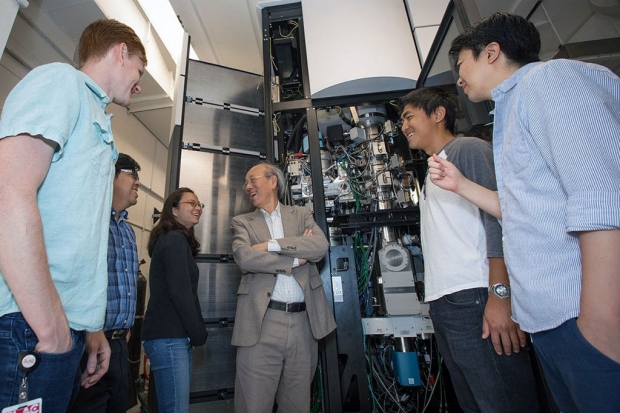
Wah Chiu and members of the cryo-EM team stand in front of a cryo-EM instrument as work nears completion on their new facility at SLAC.
Dawn Harmer/SLAC
A new facility for cryogenic electron microscopy, or cryo-EM, has opened at the Department of Energy’s SLAC National Accelerator Laboratory.
Built and operated in partnership with Stanford University, it’s equipped with four state-of-the-art instruments for cryo-EM, a technology whose rapid development over the past few years has given scientists unprecedented views of the inner workings of the cell.
The facility is the first to open as part of the Stanford-SLAC Cryo-EM Initiative, and it is one of the most advanced in the world in terms of the number of high-end instruments and level of expertise it makes available. One of the goals of the cryo-EM initiative is to get ever-more detailed 3D images of DNA, RNA, proteins, viruses, cells and the tiny biological machines within the cell.
The facility allows researchers to prepare samples, collect data at high speed and assess the quality of that data on the fly so they can make the best use of their experimental time. They can carry out experiments in person or remotely.
“This facility is the result of several years of work and planning by faculty and other leaders at SLAC and Stanford, and it’s a great example of the opportunities our partnership brings us,” said Stanford President Marc Tessier-Lavigne, PhD. “Cryo-EM has become an essential tool for research, especially in structural biology, and we’re excited that this state-of-the-art facility is finally here.”
An unprecedented view of life's machinery
Cryo-EM is a version of electron microscopy, which was invented in the 1930s. These microscopes use beams of electrons rather than light to form images of samples. Because the wavelength of an electron is much shorter than the wavelength of light, electron beams reveal much smaller things.
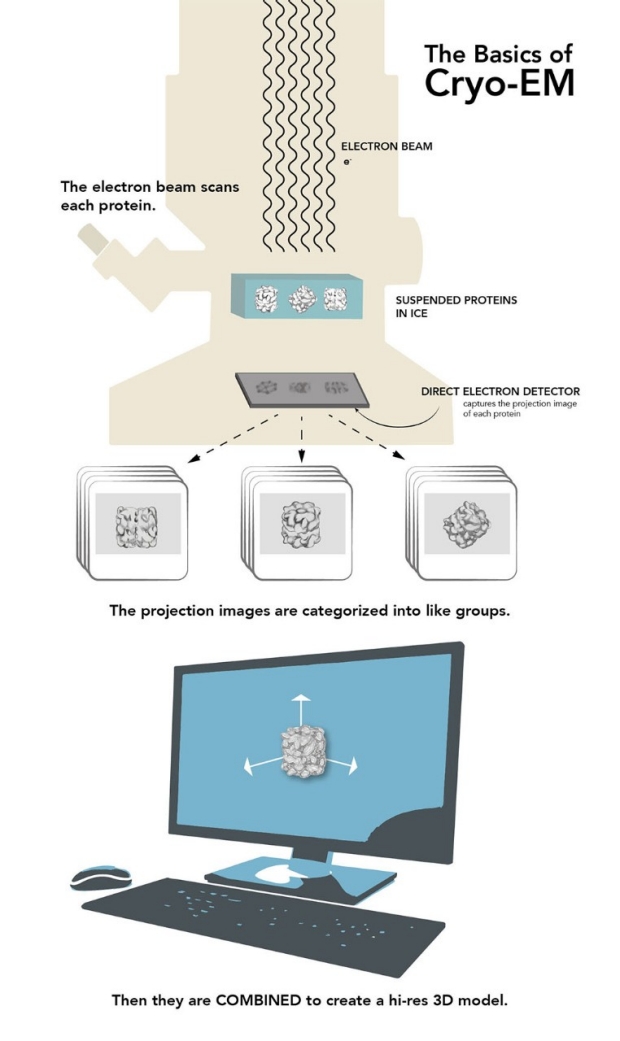
This infographic explains the basics of cryo-EM. Farrin Abbott/SLAC
In the mid-1970s, scientists came up with the idea of freezing samples to preserve the natural structure of biological specimens and reduce damage from the electron beam, and cryo-EM was born. The technology slowly evolved, and then a few years ago took a giant leap, thanks to dramatic advances in detectors and software. In 2017, three scientists were awarded the Nobel Prize in chemistry for their roles in developing cryo-EM.
Today, cryo-EM generates 3D images at nearly atomic resolution of viruses, molecules and complex biological machines inside the cell — such as the ribosomes, where proteins are synthesized. By flash-freezing these tiny things in their natural environments, scientists can see how they are built and what they do in much more detail than before, stringing thousands of images together to create stop-action movies and even taking virtual “slices” through cells, much like miniature CT scans. Meanwhile, cryo-EM instruments have become easier to use and much more accessible.
“In biology, everything is dynamic, always moving around and changing. Cryo-EM lets you capture snapshots of proteins and other biological nanomachines as they assemble, carry out their work and disassemble again,” said Wah Chiu, PhD, professor of bioengineering and of microbiology and immunology at Stanford and of photon science at SLAC. Chiu is one of two faculty members hired last year who bring decades of experience in cryo-EM research and technology development.
“A protein may multitask, changing its shape for each of its functions,” Chiu said. “From how it looks, you can determine how to modify its shape and consequently its functions — for instance, if you want to block its activity with a vaccine or develop a medication that fits into a particular pocket and triggers a response that fights disease.” He added that his own ambition is to be able to look at these biological machines at work while they’re still inside the cell, without having to remove or purify them.
Georgios Skiniotis, PhD, professor of molecular and cellular physiology and of structural biology, arrived at the School of Medicine last year from the University of Michigan. He specializes in studies of complex receptors on the cell’s outer membrane that are important targets for drug development, but whose structure and function are still poorly understood. Cryo-EM makes them much easier to study in detail.
“Many refer to it as the cryo-EM revolution, but I view it more as accelerated evolution because most of these concepts and tools have been steadily evolving for many years,” Skiniotis said. “Without any sense of exaggeration, the technology offers unprecedented imaging capabilities. Cryo-EM can be used for so many purposes.”
In battery research, for instance, scientists working with an older cryo-EM instrument at the School of Medicine recently captured the first atomic-level images of fingerlike growths called dendrites that can pierce the barrier between battery compartments and trigger short circuits or fires.
Technology and the human touch
One big advantage of locating the new facility at SLAC is the lab’s long history of technology development, including computing and data analysis, said Soichi Wakatsuki, PhD, a professor of photon science at SLAC and of structural biology at the School of Medicine, who leads the lab’s Biosciences Division.
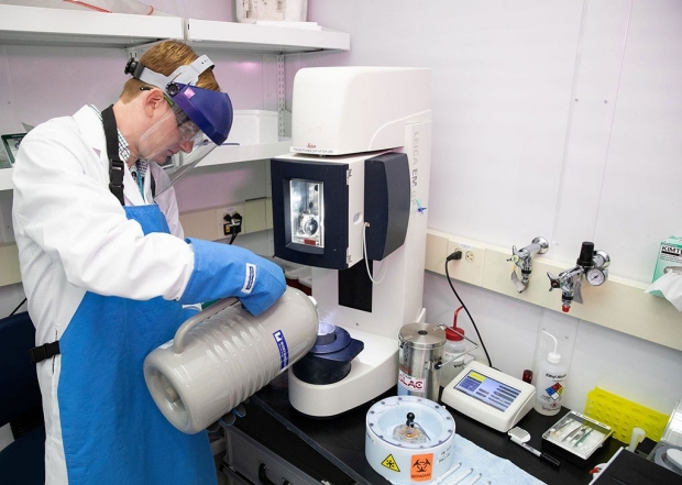
Graduate student Patrick Mitchell pours liquid nitrogen into a device used to freeze samples prior to study.
Andy Freeberg/SLAC
“Adding cryo-EM to our established X-ray approaches will help us build the fundamental understanding that researchers need to design biofuels and biomaterials, predict how carbon cycles through geological and living systems and tease out the complicated interactions of fungi and other organisms in the root zones of plants, which are important for understanding natural ecosystems as well as improving soil fertility,” Wakatsuki said.
Both Chiu and Skiniotis have been collaborating with Stanford scientists on research projects over the years, and that’s even easier now that they — and the cryo-EM facility — are close by, Chiu said.
“I think the environment of physicists, engineers, computational scientists and chemists around the SLAC and Stanford campuses can inspire us to think differently,” he said. “There’s no doubt in my mind that this will lead to new approaches in specimen preparation, data collection, image processing — in the whole pipeline.”
Chiu brings with him two National Institutes of Health research programs he started while at Baylor College of Medicine. One is a regional consortium for cryo-EM data collection. It makes instruments available to cryo-EM scientists across the United States who would not otherwise have access to the technology. The other, a national center for 3D electron microscopy of macromolecules, is focused on cryo-EM technology development that is driven by the biomedical projects of collaborators around the globe, as well as on training and serving the global community of cryo-EM researchers.
Path to the Stanford-SLAC Cryo-EM Initiative
Roger Kornberg, PhD, professor of structural biology and 2006 Nobel laureate in chemistry, has used cryo-EM for many years in research on how the cell carries out instructions in DNA, and has advocated for expansion in this area. Investments from the School of Medicine initiated the drive to expand access to this technology for the Stanford research community and, along with contributions from the President’s Office and SLAC, enabled the hiring of Skiniotis and Chiu.
“We were extremely pleased to have received this tremendous support,” said William Weis, PhD, professor and chair of structural biology. Weis and Axel Brunger, PhD, professor of molecular and cellular physiology and of neurology and neurological sciences, and Norbert Pelc, ScD, professor of bioengineering, played a major part in recruiting the new faculty members
“This is a wonderful achievement for the whole interdisciplinary team that has been working diligently to develop this program,” said Pelc, the former chair of bioengineering. “It is also a great opportunity for scientists from SLAC, Stanford and other institutions who will have access to this leading-edge technology. We all will benefit from the discoveries they will make.”
Brunger, the former chair of molecular and cellular physiology, added, “Cryo-EM now enables the study of biomolecules in their native environment, such as in membranes, and ultimately, to study them in their cellular context. Further developments in technology are conceivable and could revolutionize the fields of cell biology and molecular neuroscience.”
About Stanford Medicine
Stanford Medicine is an integrated academic health system comprising the Stanford School of Medicine and adult and pediatric health care delivery systems. Together, they harness the full potential of biomedicine through collaborative research, education and clinical care for patients. For more information, please visit med.stanford.edu.

