This spring Stanford Health Care began using the Mohs technique for melanoma in situ, which is less expensive than the traditional surgical approach, creates a smaller wound and reduces the cancer’s rate of recurrence.
June 16, 2016 - By Sara Wykes
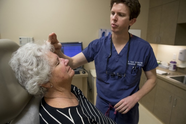
Edyth Ledbetter attends a follow-up appointment with dermatologic surgeon Tyler Hollmig, who removed melanoma in situ from her nose using a technique called Mohs surgery.
Norbert von der Groeben
Edyth Ledbetter had had enough common skin cancers removed from her face to recognize that the dark, fast-spreading spot on the side of her nose was something different.
She was right. Three months ago, Ledbetter became one of the estimated 76,000 people in the United States diagnosed with melanoma in 2016. Melanoma is the most lethal of all skin cancers, and its incidence is rising steadily while its mortality rate remains stubbornly stable. Even when melanoma is in situ — an early stage during which the cancer has not yet grown beyond the outer layer of skin — removing it is often a multistage process that takes a few days as patients and doctors wait for tissue analysis. Without the finesse of a layer-by-layer surgery called Mohs, which is used universally for other forms of skin cancer, removal of early-stage melanoma can produce large wounds and disfiguring scars.
This spring, just in time for Ledbetter’s surgery, Stanford Health Care began using the Mohs technique for melanoma in situ. This outpatient surgery procedure is less expensive than the traditional surgical approach, creates a smaller wound and reduces the cancer’s rate of recurrence. A melanoma in situ can be removed, and reconstructive surgery completed, in one day.
Preparing melanoma tissue samples
Using Mohs to treat melanoma in situ was not possible until researchers figured out how to prepare tissue samples of the cancer. Unlike more common forms of skin cancer, such as basal cell or squamous cell carcinoma, melanoma’s cells are difficult to visualize with the standard dyes used to prepare slides for analysis. About 15 years ago, researchers finally developed an immunohistochemistry stain for melanoma, tuned to the cancer’s particular biomarkers. That discovery made melanoma visible, but initially it was only used for formalin slides, which take about 12 hours to prepare, not the quicker, frozen-section slides used in Mohs surgery.
Doctors wanted to do with melanoma what they could do with other skin cancers: remove a tissue sample that could be stained, frozen and slide-ready in less than an hour. Clinicians were hesitant to use the new stain in the quick-processing preparation until studies showed it was as accurate as the previous formalin-slide-based, multiday analysis.
That evidence was published last year in the Journal of the American Academy of Dermatology. “That study added to the growing body of evidence that shows the incredible usefulness of this immunohistochemical marker in frozen-section analysis in Mohs for in situ melanoma,” said surgeon Tyler Hollmig, MD, clinical assistant professor of dermatology at the School of Medicine. “We are incredibly excited about those results and about Stanford Health Care’s ability to now offer this new technique to our patients.”

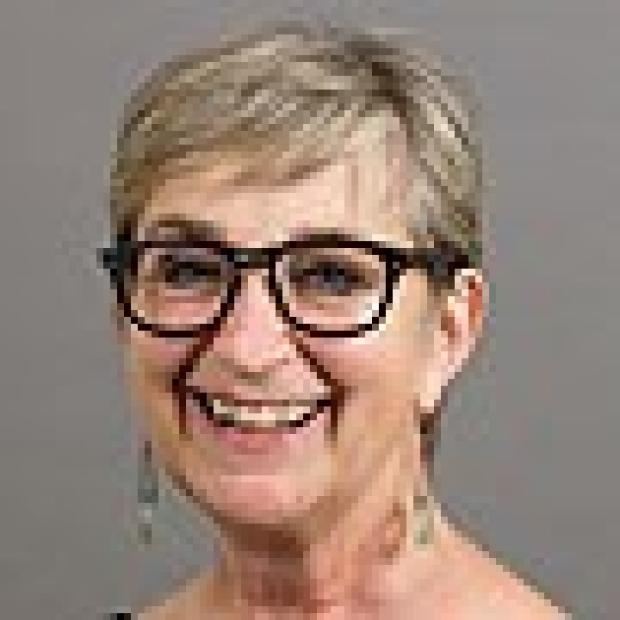
Ledbetter had the surgical wound on her nose patched with a flap from her forehead. She has said that her friends can't tell she's had surgery.
Norbert von der Groeben
Proceeding carefully
Hollmig and his Stanford Medicine colleagues have proceeded carefully because the new processing and analysis are technically difficult. “We wanted to do things the right way,” he said. Stanford Health Care’s dermatopathology service became a crucial partner for the advanced training needed for the new procedure.
The procedure is based on a technique developed by Wisconsin surgeon Frederic Mohs in the 1930s. Mohs surgery begins with the removal, like a plug, of the most visible center of the cancer. Then, the surgeon will remove tissue around the center, in layers about 3 millimeters thick. That tissue is sliced into micron-thin pieces, stained, frozen and examined within minutes. The incremental removal of tissue means that no more will be removed than is necessary. When the cancer is near the eyelid, for instance, the precision of Mohs keeps wounds and scars, or reconstructive surgery, to a minimum.
Hollmig said he and fellow dermatologic surgeon Sumaira Aasi, MD, a clinical professor of dermatology at Stanford, expect to use the procedure to treat the increasing number of people diagnosed with melanoma. The new technique is well-suited for patients whose melanoma is in areas where sparing tissue is most critical — the head, neck, hands, feet and genitalia. With speedy analysis of cancerous tissue now available, patients can have a tumor removed and any needed wound repair completed in one day.
Ledbetter, who lives in Lodi, said she appreciated that. Her melanoma had spread through half of one side of her nose. After removing the cancerous tissue, Hollmig built Ledbetter a new nose with a procedure called a paramedium forehead flap. The flap is a section of tissue about a half-inch deep and 5 inches long surgically separated away from the forehead. One edge remains attached to the forehead, just above the eyebrow; the rectangular remainder is turned down toward the nose to cover the area where cancer has been removed.
In about three weeks, the flap has healed over the nasal wound. Then it can be separated completely from the forehead, and its top edge sewn to the top of the nose. Although the flap procedure is complicated, the one-day process enabled Ledbetter to arrive at the Outpatient Center in Redwood City at 8 a.m., Feb. 17, and head home at 4:45 p.m. Two months later, her healing was not considered complete, but she has boasted that her friends “can’t tell I’ve had surgery.”
“Hearing that from a patient is my best reward,” Hollmig said.
-
 Sara WykesSara Wykes is a writer for the Stanford Hospital & Clinics communications office. Email her at swykes@stanfordhealthcare.org.
About Stanford Medicine
Stanford Medicine is an integrated academic health system comprising the Stanford School of Medicine and adult and pediatric health care delivery systems. Together, they harness the full potential of biomedicine through collaborative research, education and clinical care for patients. For more information, please visit med.stanford.edu.

