Researchers have developed a new way to use atomic force microscopy to rapidly measure the mechanical properties of cells, an advance that could pave the way for better understanding immune disorders and cancer.
December 16, 2015 - By Andrew Myers
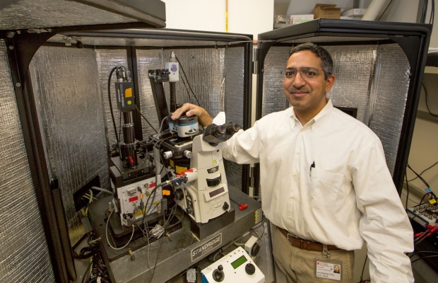
Manish Butte and his colleagues developed a way to probe and get detailed measurements of living cells through an advancement in atomic force microscopy, a technology invented at Stanford in 1986.
Norbert von der Groeben
In his role as a pediatrician, Manish Butte, MD, PhD, will often push and prod a patient’s abdomen, feeling for abnormalities — a swollen spleen, a hardened lymph node or an unusual lump in the intestines or liver. There are still some things that can only be gleaned by touch, and Butte believes this notion applies to individual cells as well.
Yet researchers’ ability to probe and measure the features of living cells has been almost nonexistent. Recently, a team of Stanford scientists and engineers set out to right that imbalance with a new technique for rapidly mapping cells. They succeeded by engineering a major advancement in a technology known as atomic force microscopy, or AFM, which itself was invented at Stanford in 1986.
A paper describing the work was published online Nov. 11 in ACS Nano. Butte, an assistant professor of pediatric immunology, is the senior author. Lead authorship is shared by Andrew Wang, PhD, a former postdoctoral scholar in Butte’s lab, and Karthik Vijayraghavan, PhD, who was a graduate student and member of the microphotonics lab led by Olav Solgaard, PhD, a professor of electrical engineering.
“What a cell feels like — its mechanical properties that affect how it makes contact with other cells and tissues — is much more important than what it looks like, but the technology just wasn’t there to allow us to examine it,” Butte said. “There is a lot to be learned from studying the mechanics of a cell and its structures just beneath the surface.”
The way Butte and his colleagues use AFM to measure the mechanical properties of cells is akin to the way a builder taps her knuckles along a drywall, listening for the change in pitch that will tell her a wooden stud is on the other side. When an AFM probe taps the surface of a cell, it vibrates, and the pattern of these vibrations, like the sound waves reflecting from the stud, gives mechanical information about the structures of the cell being touched.
However, existing AFM probes are relatively large and, as a result, insensitive to high frequencies, which communicate much of the key information about a cell’s innards. The Stanford team’s device couples a very small probe with a traditional one. This assembly allows the device to sense faster oscillations than conventional devices and, accordingly, to take more detailed and much faster measurements.
“The main difference between this and previous atomic force microscopes is that we are able to measure the impact of the probe on the cell very fast and get specific readings, whereas typical AFMs simply provide an average. This allows us to accurately measure some very soft materials for the first time,” said Solgaard, who also is a co-author of the paper.
Current probes measure cellular stiffness by tapping against the cell around one or two times per second — the fastest that the large probes can make measurements. The small probe, however, can make detailed measurements easily at five to 10,000 taps per second because of its sensitivity. He likened the leap in sensitivity to the difference between driving a Cadillac Escalade down the road and pushing a Hot Wheels toy car along the same surface: “The small Hot Wheels will feel every little bump so much more than the large Cadillac.”
‘Beautiful solution’
AFMs measure movement of the probe by bouncing a laser off its tip. As the tip moves up and down, the laser is reflected. The Stanford invention couples the small probe with the large one by means of a fork-shaped structure called an interferometric grating. The grating produces a diffraction pattern based on the movements of the small probe, and allows the AFM to conveniently capture its measurements.
What a cell feels like — its mechanical properties that affect how it makes contact with other cells and tissues — is much more important than what it looks like.
“Our tip actually produces a second signal, and that is what allows us to get much greater detail. From an engineering standpoint, it’s an extremely simple, beautiful solution,” Solgaard said, referring to the diffracted signals from the grating.
Best of all, the team’s device can be directly attached to existing AFMs, potentially saving millions of dollars on new equipment that could otherwise be spent on research. A new AFM can run as much as $500,000, according to Solgaard.
The objective is the cellular equivalent of Butte pressing a child’s abdomen.
“We want to study cell stiffness to understand what is beneath the surface and how cells are structured,” Wang said.
As a demonstration, the team measured a section of a red blood cell, making approximately 4 million total measurements in about 10 minutes — all without damaging the delicate cellular exterior.
“The same measurements would have taken more than a month to complete using conventional atomic force microscopes,” said Vijayraghavan. The technology is so fast that the team was able to create a series of time-lapse images of a living cell, each taken just seven minutes apart, a previously unimaginable pace.
Potential applications
The practical applications of the device range from basic scientific understanding of cellular structure to immunology and oncology. Scientific understanding of the mechanical forces at play in cells is so lacking that the field — now being called mechanobiology — is really in its infancy, according to Butte.
The same measurements would have taken more than a month to complete using conventional atomic force microscopes.
The mechanical forces in the body can come from tissues, which range in stiffness from softest brain matter to stiffest bones, from gravity, and even from the pushing and pulling movements of other cells. Cancer cells make their environment mechanically rigid by secreting chemicals that stiffen up the extracellular matrix. Cancer cells likewise interpret the mechanical forces of a tissue to make decisions about growth and metastasis. Surprising feedback loops like this also appear to occur for stem cells in the bone marrow and during embryonic development. How immune cells interpret mechanical forces is still totally unknown.
“The lowest-hanging fruit is cancer. Cancers are often stiffer than normal, healthy tissues and we can use that knowledge to diagnose disease. But first, you have to have good data, which our device provides,” Wang said. He has already used an early form of the new Stanford probe in pilot work on breast cancer specimens taken from mastectomies.
For his part, Butte plans to use fast AFM to study the immune system. He hopes to explore why otherwise disease-fighting T cells often remain dormant once inside a tumor. He theorizes that the mechanical stiffness of the tumorous tissue may be preventing T cells from freely making contact with cancer cells and from triggering their cancer-fighting functions. In essence, the tumor may be too crowded for the T cells to work. On the other end of the stiffness gamut, he believes that the soft mechanical properties of chronically inflamed or infected tissues provoke the immune system into over-activity, like autoimmunity.
It is a theory no one has yet explored due to technical barriers, which the fast AFM could overcome. Butte’s lab has begun a broad effort to link mechanical forces with immune responses at the molecular, cellular and tissue scales. “There is so much we don’t know about the mechanical properties of various cell types and diseased tissues. Almost nothing, in fact,” Butte said. “The first step is to probe. Now, we can do that.”
The work was funded by the National Institutes of Health (grants K08AI079268 and R01GM110482), the National Science Foundation, the Stanford Center for Probing the Nanoscale, the Stanford Child Health Research Institute and Stanford Bio-X.
The departments of Pediatrics and of Electrical Engineering also supported the work.
About Stanford Medicine
Stanford Medicine is an integrated academic health system comprising the Stanford School of Medicine and adult and pediatric health care delivery systems. Together, they harness the full potential of biomedicine through collaborative research, education and clinical care for patients. For more information, please visit med.stanford.edu.

