February 16, 2014 - By Krista Conger

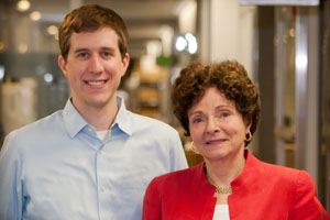
Benjamin Cosgrove, Helen Blau and their colleagues have identified a process by which older muscle stem cell populations can be rejuvenated to function like younger cells.
Researchers at the Stanford University School of Medicine have pinpointed why normal aging is accompanied by a diminished ability to regain strength and mobility after muscle injury: Over time, stem cells within muscle tissues dedicated to repairing damage become less able to generate new muscle fibers and struggle to self-renew.
“In the past, it’s been thought that muscle stem cells themselves don’t change with age, and that any loss of function is primarily due to external factors in the cells’ environment,” said Helen Blau, PhD, the Donald and Delia B. Baxter Foundation Professor. “However, when we isolated stem cells from older mice, we found that they exhibit profound changes with age. In fact, two-thirds of the cells are dysfunctional when compared to those from younger mice, and the defect persists even when transplanted into young muscles.”
Blau and her colleagues also identified for the first time a process by which the older muscle stem cell populations can be rejuvenated to function like younger cells. “Our findings identify a defect inherent to old muscle stem cells,” she said. “Most exciting is that we also discovered a way to overcome the defect. As a result, we have a new therapeutic target that could one day be used to help elderly human patients repair muscle damage.”
Blau, a professor of microbiology and immunology and director of Stanford’s Baxter Laboratory for Stem Cell Biology, is the senior author of a paper describing the research, published online Feb. 16 in Nature Medicine. Postdoctoral scholar Benjamin Cosgrove, PhD, and former postdoctoral scholar Penney Gilbert, PhD, now an assistant professor at the University of Toronto, are the lead authors.
The researchers found that many muscle stem cells isolated from mice that were 2 years old, equivalent to about 80 years of human life, exhibited elevated levels of activity in a biological cascade called the p38 MAP kinase pathway. This pathway impedes the proliferation of the stem cells and encourages them to instead become non-stem, muscle progenitor cells. As a result, although many of the old stem cells divide in a dish, the resulting colonies are very small and do not contain many stem cells.
Using a drug to block this p38 MAP kinase pathway in old stem cells (while also growing them on a specialized matrix called hydrogel) allowed them to divide rapidly in the laboratory and make a large number of potent new stem cells that can robustly repair muscle damage, Blau said.
“Aging is a stochastic but cumulative process,” Cosgrove said. “We’ve now shown that muscle stem cells progressively lose their stem cell function during aging. This treatment does not turn the clock back on dysfunctional stem cells in the aged population. Rather, it stimulates stem cells from old muscle tissues that are still functional to begin dividing and self-renew.”
The researchers found that, when transplanted back into the animal, the treated stem cells migrate to their natural niches and provide a long-lasting stem cell reserve to contribute to repeated demands for muscle repair.
“In mice, we can take cells from an old animal, treat them for seven days — during which time their numbers expand dramatically, as much as 60-fold — and then return them to injured muscles in old animals to facilitate their repair,” Blau said.
In 2010, Blau’s laboratory published a study in Science showing that muscle stem cells grown on soft hydrogel maintain their “stemness” in culture. In contrast, muscle stem cells grown on hard plastic tissue culture plates, the standard way to cultivate cells in the laboratory, quickly differentiate into more-specialized, but less therapeutically useful, muscle progenitor cells. The difference is likely due to the fact that soft hydrogel is more similar than rigid plastic to the muscle tissue environment in which the stem cells are naturally found.
In the current study, the researchers found that targeting the p38 MAP kinase to induce the rapid expansion of the remaining functional stem cells from old mice required the soft hydrogel substrate. “The drug plus hydrogel boosts the small clones so that they undergo a burst of self-renewing divisions,” Gilbert said. Thus, rejuvenation of the population is contingent on the synergy between biophysical and biochemical cues.
Finally, the researchers tested the ability of the rejuvenated old muscle stem cell population to repair muscle injury and restore strength in 2-year-old recipient mice. They teamed up with co-author Scott Delp, PhD, the James H. Clark Professor in the School of Engineering, who has designed a novel way to measure muscle strength in animals that had muscle injuries and then underwent the stem cell therapy.
“We were able to show that transplantation of the old treated muscle stem cell population repaired the damage and restored strength to injured muscles of old mice,” Cosgrove said. “Two months after transplantation, these muscles exhibited forces equivalent to young, uninjured muscles. This was the most encouraging finding of all.”
The researchers plan to continue their research to learn whether this technique could be used in humans. “If we could isolate the stem cells from an elderly person, expose them in culture to the proper conditions to rejuvenate them and transfer them back into a site of muscle injury, we may be able to use the person’s own cells to aid recovery from trauma or to prevent localized muscle atrophy and weakness due to broken bones,” Blau said. “This really opens a whole new avenue to enhance the repair of specific muscles in the elderly, especially after an injury. Our data pave the way for such a stem cell therapy.”
Other Stanford authors of the study include postdoctoral scholar Ermelinda Porpiglia, PhD; instructor Foteini Mourkioti, PhD; undergraduate student Steven Lee; senior research scientist Stephane Corbel, PhD; and medical resident Michael Llewellyn, MD, PhD.
The research was supported by the National Institutes of Health (grants R25CA118681, K99AG042491, T32CA009151, K99AR061465, U01HL100397, U01HL099997, R01AG020961, R01HL096113 and R01AG009521), the California Institute for Regenerative Medicine and the Baxter Foundation.
Information about Stanford’s Department of Microbiology and Immunology, which also supported the work, is available at http://microimmuno.stanford.edu.
About Stanford Medicine
Stanford Medicine is an integrated academic health system comprising the Stanford School of Medicine and adult and pediatric health care delivery systems. Together, they harness the full potential of biomedicine through collaborative research, education and clinical care for patients. For more information, please visit med.stanford.edu.