February 22, 2010 - By Erin Digitale
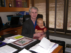
Frank Hanley holds Audrey Walker after giving her a new lease on life.
Frank Hanley, MD, the pioneer of “unifocalization” surgery to repair complex cardiac defects in kids, is world-known for tackling cases that surgeons in places like Israel, Belgium and Australia would not touch. Now, the surgeon-researcher at Lucile Packard Children’s Hospital is attacking the problem that troubles him most: How to grow durable replacement valves for tiny, defective hearts.
The results could be revolutionary. “If we could form a heart valve with a patient’s own tissue that would grow and heal itself, that would be a huge advance for kids and even adults,” Hanley said of his current lab research, explaining that today’s prosthetic or cadaver replacements wear out and create the need for additional surgeries. Together with regenerative medicine experts, Hanley and his team are trying to determine ways to engineer fully functional, natural-tissue heart valves that grow with the patient and also self-repair.
No one understands the potential impact better than the families of the more than 500 children whose lives Hanley has saved with unifocalization, such as Jim and Heather Walker of San Jose. Their daughter Audrey was born without the vessels carrying blood to her lungs, she had a hole in her heart and she was missing a heart valve. These complex and deadly defects limited her lungs’ ability to supply oxygen.
The solution? Hanley’s “let’s fix everything at once” unifocalization. It’s a grueling, 12- to 14-hour surgical epic that, before Hanley began developing the technique in the early 1990s, other surgeons thought just couldn’t be done. Instead, these children faced the trauma and danger of three heart and lung surgeries spread over years of childhood.
“Unifocalization is unbelievably demanding,” said Stephen Roth, MD, director of the cardiac intensive care unit at Packard Children’s. “And no one is better than Dr. Hanley at performing such a meticulous and detailed operation.”
For instance, Hanley explained a lung fix. “The blood vessels from the heart to the lungs are supposed to look like an oak tree: The pulmonary artery ‘trunk’ sends a big branch to each lung, and these vessels branch again and again to reach the lung’s air sacs,” said Hanley, a professor of pediatric cardiothoracic surgery at the School of Medicine and the Lawrence Crowley, MD, Professor in Child Health.
In children like Audrey, the lung blood vessels look as though “someone took a buzz saw to a normal-sized oak tree, cut off four or five of the first big branches and scattered them around the field. What we do is the equivalent of gathering those scattered branches back together and try to make an oak tree,” he said.
Before Hanley came along, other surgeons would perform a separate operation on the blood vessels in each lung, then a third surgery on the heart. Though it requires incredible stamina from the surgeon, Hanley found that unifocalization’s 3-in-1 approach greatly improved patients’ outcomes. In Audrey’s operating room marathon, for instance, Hanley didn’t just dissect all the abnormal blood vessels leading to her lungs and painstakingly reshape them into a two-branched pulmonary artery. He also repaired all the other defects, such as closing the hole in the heart and replacing the missing heart valves with those from a cadaver—the step that is the target of his new research.
Building personalized heart valves
“Children outgrow T-shirts and shoes—and kids can outgrow the valves we use, or the valves wear out,” Hanley said. “Unfortunately, patients like Audrey still may have to come back and get these valves replaced.”
The solution? Forming new heart valves from the patient’s own tissue. That’s why Hanley and his regenerative medicine collaborators, senior research scientist Kirk Riemer, PhD, and Michael Longaker, MD, professor of surgery and the director of children’s surgical research at Packard Children’s, are studying what goes on inside normal valves, the paper-thin leaves of tissue that prevent blood from backwashing on its trip through the heart.
“The heart contracts 40 million times a year,” said Riemer. “The valves are very busy pieces of tissue. They flap like flags in a continuous windstorm.
“We think if the valve starts to lose cells, a small breach can bring about series of reactions that manifest as a big problem, like the tiles on the space shuttle,” Riemer added. Normally, the body stops this big problem before it starts; to find out how this happens, the team’s research will have to detect miniscule changes in chemical signals sent out by valve cells, then figure out how such signals work.
Ultimately, the researchers hope to program a patient’s own stem cells to grow into functioning heart valves that last a lifetime. “The regenerative principles we are in the process of discovering could create new valve and vascular graft durability for both kids and adults,” said Riemer.
“Unifocalization works, but we are still looking for a way to avoid a later valve replacement surgery,” said Hanley. “The ultimate goal is to operate on a patient like Audrey once and say, ‘she’s cured.’ It’s the next great frontier in our work, and we’re hoping this research can make it a reality.”
-
 Erin DigitaleErin Digitale is the pediatrics science writer for the medical school’s Office of Communication & Public Affairs.
About Stanford Medicine
Stanford Medicine is an integrated academic health system comprising the Stanford School of Medicine and adult and pediatric health care delivery systems. Together, they harness the full potential of biomedicine through collaborative research, education and clinical care for patients. For more information, please visit med.stanford.edu.

