Using machine learning to identify individual variations in the primate retina
April 12, 2022 - By Dian Le

Using large-scale electrode recordings of the primate retina, Stanford researchers identified quantitative measures of individual variability. Their discoveries contribute to their development of an artificial retina for humans.
What does the eye tell the brain? This question drives the Chichilnisky lab’s work. Now, lab researchers have found individual differences in how primate retinas process light stimuli and transmit visual signals to the brain.

Nishal Shah, PhD
In a recent study published in Neuron, Stanford researchers developed a new way to analyze how the neural code of the retina varies across individuals. They applied this method to a unique dataset – over a decade of large-scale recordings from macaque retinas, the most relevant model for the human retina. Nishal Shah, PhD, a former graduate student (and now a postdoctoral scholar in the Henderson and Shenoy lab) is the study's lead author.
In the study, the research team recorded electrical activity as retinal cells fired in response to visual stimuli. Shah described the study as a listening exercise. "The retina is in its natural operating mode – looking at light – and we are just overhearing the activity," he said.
The researchers examined the neural code in approximately 100 macaque retinas and quantified the individual variation using machine learning methods.
"We know that every individual has a unique nervous system that determines their perception and behavior," said Shah. "But until now no studies have been able to quantitatively capture or model this variation."
The results revealed "surprising and systematic differences in the visual processing in male and female retinas," according to Shah. The research team found that male retinas exhibited higher firing rates and faster processing than female retinas.
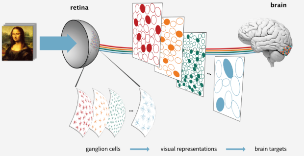
The Chichilnisky lab studies how light stimuli are encoded by ganglion cells in the retina. Ganglion cells react to light entering the eye and transmit these signals toward specific areas of the brain. (Source)
The results of the study, according to the research team, further contribute to the development of a retinal implant by the Artificial Retina Project. Led by the Chichilnisky lab, the Artificial Retina Project is a collaborative effort involving neuroscientists, electrical engineers, and a retinal surgeon. Their work aims to create technology that can restore sight to people blinded by retinal degenerative disease.
In the full Q&A with the study authors below, Drs. Shah and Chichilnisky shared insights from their latest publication.
On their paper's significance:
Our paper captures for the first time how the neural circuitry of the visual system varies across individuals. We know that every individual has a unique nervous system that determines their perception and behavior. But until now no studies have been able to quantitatively capture or model this variation.
We discovered a systematic variation in visual processing in the primate retina using artificial neural networks and a unique dataset of nearly 100 large-scale recordings of roughly 500 neurons from each retina. In addition to showing how vision varies across individuals, the work revealed surprising and systematic differences in the visual processing in male and female retinas.
How will your study findings impact your clinical goals?
Understanding the variation in visual processing between individuals will permit us to customize the development of novel electronic implants for treating blindness. We envision doing so by using behavioral tasks to identify the unique aspects of visual processing in the implant recipient, and then adjusting the implant’s function in software to best fit the recipient’s needs, using the quantitative methods developed in this paper.
We also expect that the methods we developed to understand the natural variation of visual processing across individuals will aid in the diagnosis of visual deficits that are outside the range of normal individual variation.
How does your research fit into the bigger scope of the Artificial Retina Project?
Our lab has focused on understanding visual processing in the retina, using unique large-scale recording techniques to probe the patterns of electrical activity in many types of retinal neurons that convey visual information to the brain.
We had previously noticed important variations in the hundreds of recordings we had performed, but we had no systematic understanding of them. We also didn’t know how this variation would affect the collaborative effort that we lead, the Stanford Artificial Retina project, which is developing an electronic retinal implant to restore vision to people blinded by retinal degeneration.
This new work therefore ties together many years of basic research and makes it possible for us to harness this unique data set for greater clinical relevance of the basic research and the implant development.
Read the full paper in Neuron.
Part of this work was performed while Dr. Shah was a Student Researcher at Google Brain.