PET in a Nutshell
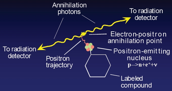
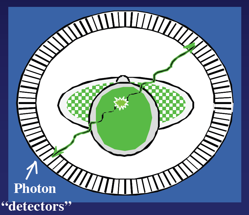
Positron emission tomography (PET) is a non-invasive biomedical imaging technology that shows promise for accurate (sensitive and specific) identification of disease due to its potential to sense, visualize, and quantified increased molecular changes in diseased compared to healthy tissue well before structural or physiological changes occur. PET uses a radioactive “molecular” contrast agent (a.k.a. the “probe”) that preferentially accumulates in diseased cells. Each radioactive decay yields two oppositely directed high-energy [511 kilo-electron-Volts (keV)] photons that are emitted as a result of positron annihilation with atomic electrons in the tissue. The PET system is typically configured as a ring of annihilation photon detectors. The detectors typically comprise >2 cm thick inorganic crystals to stop the highly penetrating photons. The detector ring collects millions of these two-photon events emitted from the body, measures spatial, energy, and temporal information for each photon detected, and, using tomographic image reconstruction methods, processes this information to reconstruct three-dimensional images of the biodistribution of the molecular contrast agent, thereby enabling the localization, quantification, and characterization of the underlying disease.
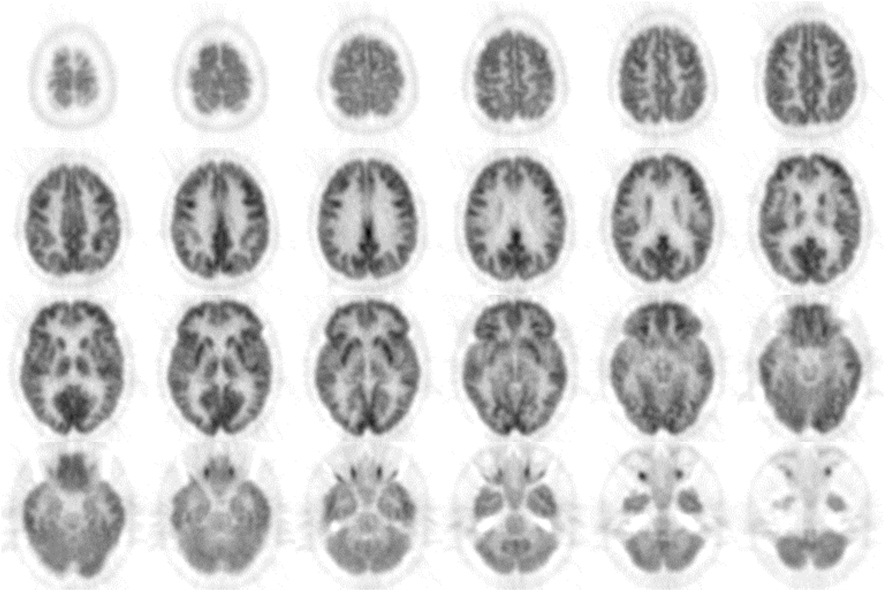
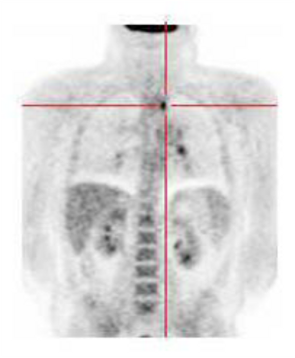
PET image examples:
Transaxial slices through a human brain using the tracer
18F-fluorodeoxyglucose (FDG)
Transaxial and Coronal slice through the whole body (FDG)

Coronal slides through a rat (FDG)
