Richard M. Lucas Center for Imaging
Location
Lucas Research MRI
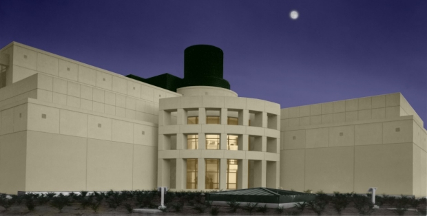
Richard M. Lucas Center for Imaging
Department of Radiology
Stanford University - School of Medicine
1201 Welch Road
Stanford, CA 94305
Who We Are
We envision that the Lucas Center will remain the creative environment for unprecedented interdisciplinary research illuminating our understanding of human physiology and lighting the way for revolutionary advances in the diagnosis and treatment of human diseases. Our mission is to advance imaging in healthcare at Stanford through technology creation and development, translational research and education.
The Lucas Center opened in July 1992 as one of the few centers in the world with major centralized resources devoted to research in magnetic resonance spectroscopy (MRS) and imaging (MRI). The Center provides office and laboratory facilities for full-time faculty members and their team of scientific staff, postdoctoral fellows and students. We actively support collaborative and original research through building upon a long-standing and very close working relationship between the faculty and students of RSL, the Clinical Radiology Department, and members of the Electrical Engineering Department often having joint seminars, journal clubs and study groups. In addition, faculty members from all groups are joint advisors to many students and have many federally funded and industry-funded collaborative research programs in place.
Our facilities are available to Stanford and non-Stanford researchers by arrangement with the Center Administrator, Jessie Leong; use is billed on a per-hour basis.
For research studies that require the use of one of the whole body magnets at the Lucas Center, please contact the Magnet Manager, Karla Epperson. The Magnet Manager or MR Research Technologists trains individual researchers in magnet safety and scanner operations. Unsponsored research projects must be pre-approved by the Research Committee of the Department of Radiology. Collaborations with Lucas Center researchers are invited and encouraged.
Yamil Saenz is a California Licensed Veterinarian (DVM), with over 22 years of experience in animal research. He provides support to the investigative staff in all animal model protocols within Radiology and for all other departments conducting research studies at the Lucas Center advanced imaging, MSLS and the Grant building Zeego Lab.
Our Facilities & Resources
The Lucas Center has 37,000 square feet of space, dedicated to imaging research, and is located on the Stanford campus, one block from the School of Medicine and Stanford University Hospital. There are four GE whole-body MRI systems, specifically three 3T, and one 7T (see Facilities for details). In addition, the Lucas Center has data analysis laboratories, an electronics laboratory/machine shop, office space, and is well suited for handling the scanning of patients and normal volunteers comfortably and safely.
Computers in the Lucas Center are networked with all scanners, including all clinical scanners, and the PACS system. Adequate electronics, mechanical laboratory facilities, machine shops and support personnel are situated at the Lucas Center and elsewhere at Stanford.
The equipment listed is available at no cost to the sponsor beyond those included in our budget. Usage costs for research scanning at the Lucas Center are included in our budget justification, and research usage of clinical systems is at similar rates. All of these costs cover maintenance of the research MRI equipment.
MRI Research Scanners
The Radiology Department at Stanford currently operates a total of 4 MRI scanners plus a mock MRI scanner:
- Two 3T GE MR 750 systems at Lucas Center (research only)
- One 3T GE MR 750 PET-MR at Lucas Center (research only)
- One 7T GE MR 950 whole-body scanner at Lucas Center (research only)
- One Mock Scanner at Lucas Center
Research scanners are available for all proposed human scans. All scanners run compatible software so pulse sequences and reconstructions can easily be supported on all systems. Reconstruction servers are networked to all scanners for reconstruction and data archiving.
All 3T scanners are equipped with state-of-the-art gradient systems (at least 50 mT/m Gradients / 150 mT/m/ms slew rates) and 32 receive channels. All 3T scanners include an assortment of RF coils including quadrature and phased-array coils designed to image brain, spine, neurovascular, torso, pelvis, cardiac, knee, foot/ankle, and hand/wrist. All 3T systems all have several sizes of 16-channel “wrap” coils that are excellent for hip and knee scans using parallel imaging.
Computer Equipment
The Lucas Center also maintains appropriate computer systems for pulse sequence programming including the GE EPIC pulse sequence programming environment, Matlab, and other statistical and analytic tools.
High-performance computers are available for more advanced imaging reconstruction. A substantial infrastructure of software tools exists to help manipulate data between all MRI scanners and reconstruction servers on different local networks, with careful support for data protection.