MR Imaging Of The Meniscal Radius Tie Sheaths
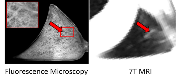
The radial tie sheaths of the meniscus are visible using fluorescence microscopy due to the natural fluorescence of collagen, as seen in the meniscus slice. The most prominent radial tie sheath structures visualized using fluorescence microscopy can also be seen using MRI (red arrows indicate aligning radial tie sheath structures).
The menisci of the tibiofemoral joint contribute to load distribution and joint stability. The radial tie sheaths of the meniscus hold together the circumferential fibers. Degradation of these radial tie sheaths is believed to predispose the meniscus to degenerative tears, putting the knee at risk of osteoarthritis. Imaging this internal structure of the meniscus is important in understanding the mechanics of the radial tie sheaths and how they degenerate. MRI protocols are being developed with the aim to image the radial tie sheaths in vivo to further understand their importance in meniscal integrity.
Publications
CONFERENCE ABSTRACTS
Black MS, Baylon G, Hargreaves B, Gold G, McWalter E, Levenston M. Non-invasive Imaging of the Radial Tie Sheaths of the Bovine Meniscus using MRI. In Proceedings of the Orthopaedic Research Society (ORS) Annual Meeting, Orlando, FL, 2016.
Black MS, Baylon EG, Varela C, Hargreaves B, Levenston M, Gold G, McWalter E. Imaging the Internal Structure of the Bovine Meniscus using MRI. In Proceedings of the 8th International Workshop on Osteoarthritis Imaging (IWOAI), Pacific Grove, CA, 2015.
Acknowledgments
The study is funded by NIH R01 AR0063643, NIH R01 EB002524, NIH K24 AR062068, and GE Healthcare.