Kinetic Modeling Of Bone Metabolism
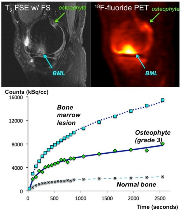
Bone marrow lesion (BML) and grade 3 osteophyte identified on 3 Tesla MRI fast spin echo images co-localize with increased uptake of [18F]-fluoride. Time activity curves showing tracer uptake over time in each region of interest is shown with the corresponding two-compartment model fit (blue).
Osteoarthritis (OA) is a complex progression of joint degeneration that manifests as tissue changes, e.g. bone marrow lesions (BMLs) and osteophytes, on MRI. To better understand the physiology that drives these changes, [18F]-fluoride is a promising PET tracer of bone remodeling within OA lesions.
Most clinical PET scans look at static accumulation of tracer after an appropriate post-injection delay. However, kinetic modeling of dynamic PET data enables quantification of rate constants that describe how the tracer is metabolized by tissue. This study investigates dynamic uptake of [18F]-fluoride in OA-related BMLs and osteophytes observed on MRI. We aim to determine whether these pathologies have different metabolic rate constants from healthy bone. These new physiological parameters may help highlight underlying mechanisms of OA pathology on MRI scans and to tell which lesions are active parts of the disease.
Publications
CONFERENCE ABSTRACTS
Fan AP, Kogan F, Patel A, Oei EH, Quon A, Gold GE. Dynamic analysis of [18F]-sodium fluoride uptake in knee osteoarthritis with PET-MRI. In Proceedings of the 24th International Society for Magnetic Resonance in Medicine (ISMRM) Annual Meeting, Singapore, Singapore, 2016.
Fan AP, Kogan F, Patel A, Oei EH, Quon A, Gold GE. Dynamic imaging of [18F]-Fluoride uptake in knee osteoarthritis with PET-MRI. In Proceedings of the OsteoArthritis Research Society International (OARSI) World Congress, Amsterdam, The Netherlands, 2016.
Acknowledgments
The study is funded by GE Healthcare, NIH EB002524, AR062068, AR0063643, and CA159992.