A Patient-Specific Mixed-Reality Visualization Tool for Thoracic Surgical Planning
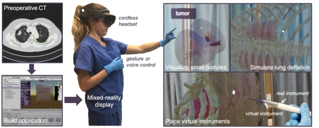
Mixed reality may help with identifying small lung lesions during minimally invasive thoracic surgery. We have developed a software application and medical image processing pipeline for the Microsoft HoloLens to incorporate patient-specific data and provide a mixed-reality tool to explore and manipulate chest anatomy in three dimensions with a custom-designed user interface featuring gesture and voice recognition. Our prototype allows the user to (1) view a patient's original preoperative imaging; (2) manipulate a 3-dimensional rendering of that patient's chest anatomy including the bronchial, osseus, and vascular structures; and (3) simulate lung deflation and surgical instrument placement.
Perkins SL*, Krajancich B*, Yang CFJ, Hargreaves BA, Daniel BL, Berry MF. A patient-specific mixed-reality visualization tool for thoracic surgical planning. Ann Thorac Surg. 2020 Jul;110(1):290-295.

 |
Bruce Daniel, MD |
 |
Brian Hargreaves, PhD |