
Jinkyung Kim
PhD

Anthony Ricci
PhD
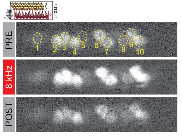
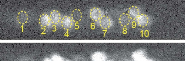
Hair cells light up as calcium movement is imaged in vivo.
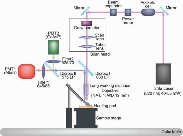
The light path of the in vivo microscope.
The cochlea remains one of the last inaccessible organs to visualize function or to perform surgical procedures. Buried deep within the temporal bone and being encased in a hard bony shell while being super sensitive to vibration and overstimulation create an almost insurmountable set of logistical problems. We are developing the surgical approach and imaging technologies needed to overcome these difficulties. Using animal models for live-cell imaging will provide unprecedented levels of detail about how the cochlea functions. Success with surgical approaches will be tested for their translational validity and may provide new access for surgeons when investigating hearing loss. The images shown here depict the custom-designed 2-photon imaging system used for viewing cellular function in vivo. The lightpath for imaging is also shown in cartoon form. Examples of multiple inner hair cells responding to sound stimulation are also presented. In, mouse we can now access the cochlea with no detectable change in hearing sensitivity.