Image Gallery
Lab showcase at the California Academy of Sciences, San Francisco
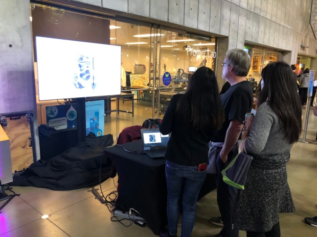
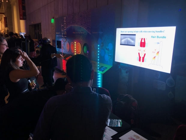
Exploring Sounds and Science
(February 20th 2020, nightlife Noise Pop Fest Showcase)


Lab Life


Scientific Pictures Gallery
Sometimes pictures are more telling than words to describe scientific experiments.
We present here some images from our research on the inner ear.
These pictures are copyright protected; please contact the lab to obtain permission for use.
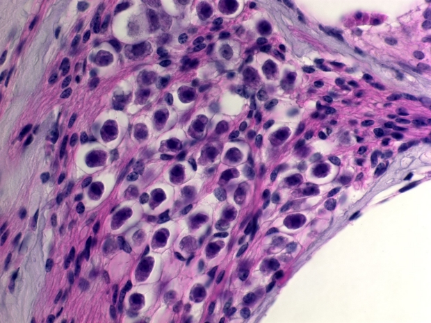
Histology

Section through the cochlea

Photograph of section trough a sensory epithelium of the vestibule sensitive to angular acceleration

Organ of Corti of a deaf animal, hair cells are absent

Organ of Corti of a normal animal, hair cells are present

Low density of cochlear neurons in a deaf animal
High density of cochlear neurons in a normal animal
Molecular Biology

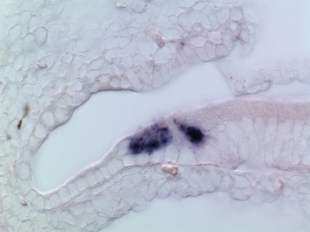
In situ hybridization on embryonic inner ear sections. The left one is using a supporting cells-specific probe, and the right one a hair cell-specific probe.
In situ hybridization on postnatal inner ear section using a hair cell-specific probe.
Mouse Genetics
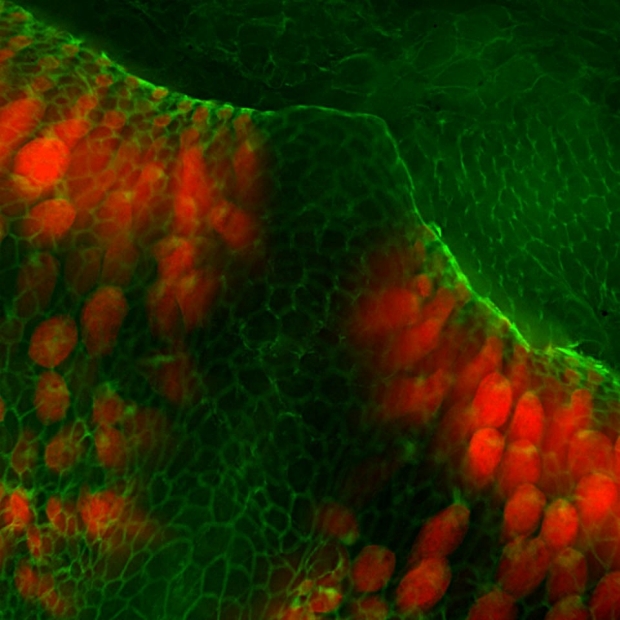
Visualization of the sensory cells of the inner ear detecting angular acceleration. We are able to detect head movement because of the ability of the vestibular part of the inner ear to detect such changes. Here we visualize the anatomy of this nervous system at the level of a semi-circular canal. The sensory cells, called hair cells, are positioned at the base of a dome. They will be stimulated upon specific angular acceleration giving us the sense of spatial positioning. Here a mouse ampulla was stained for actin (green) and the hair cells were genetically stained with TdTomato (red). This picture was finalist in the 2012 GE Healthcare Life Sciences Cell imaging competition.
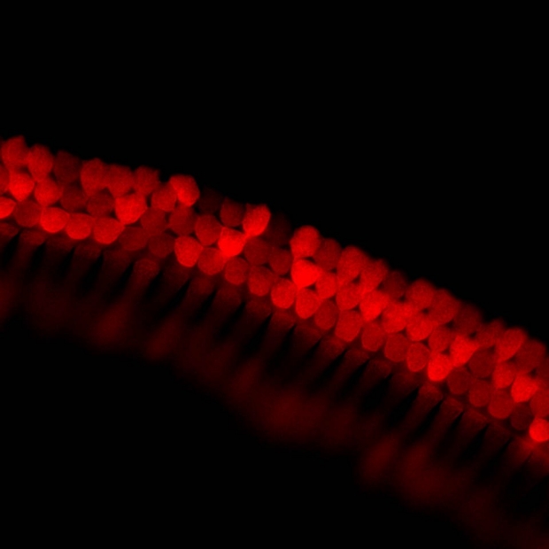
Visualization of the sensory cells of the inner ear detecting sound The cochlear part of the inner ear able us to detect sound. The sensory cells, called hair cells, are able to detect specific sound frequencies and generate an electric signal that will be sent to the brain. Hair cells are aligned along the cochlea according to their frequency sensitivity. The hair cells of the cochlea are therefore decomposing the complex sounds into individual signals, playing the inverted function of a musical instrument. Here the hair cells of a mouse cochlea were genetically stained for TdTomato (red). This picture was finalist in the 2012 GE Healthcare Life Sciences Cell imaging competition.
Fluorescence Microscopy
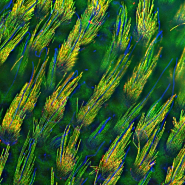
Mechanosensitive hair bundles of Vestibular Hair Cells
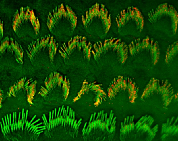
Mechanosensitive hair bundles of Cochlear Hair Cells
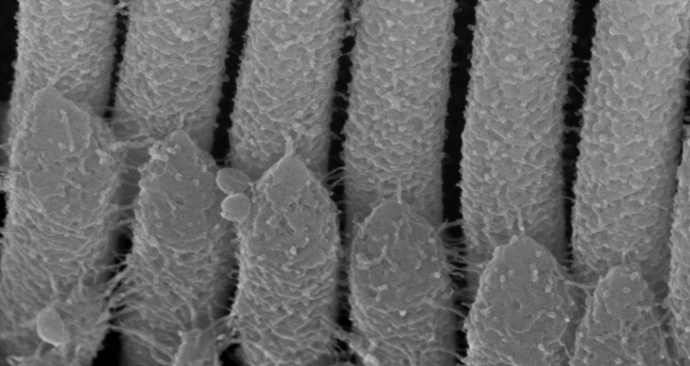
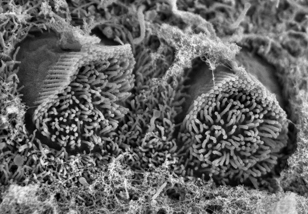
Scanning EM

Unmatured cochlear hair cell bundle and its linkage
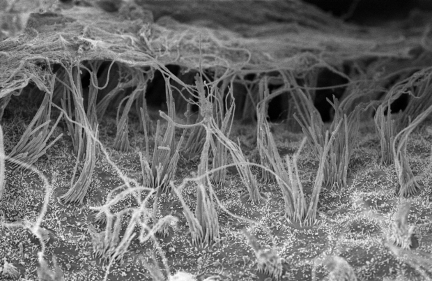
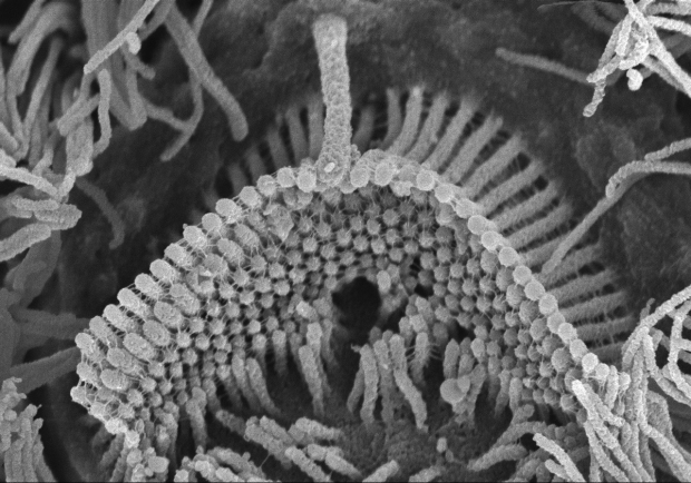
Vestibular hair cells bundles
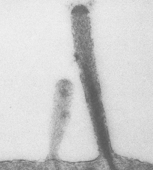
Array of tip-links from cochlear hair cells
Early stage of the cochlear hair cell bundle formation
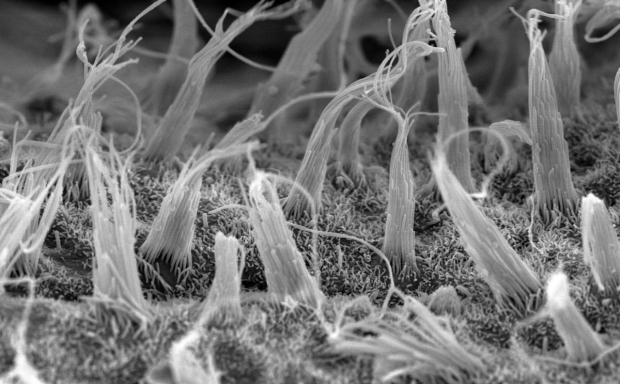
Cochlear hair cell bundle
Vestibular hair cells bundles
Transmission EM
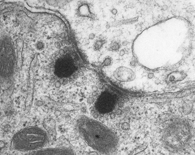
Synaptic contacts between a hair cell (bottom) and its connected neuron (top). Note the synaptic vesicles arranged along the electron-dense ribbons just in front of the synaptic cleft.
Immuno-gold labeling on a cochlear hair cell sagittal section (Grillet, 2009, Neuron).
Bioinformatics

With the Bioinformatics we can model mutations leading to deafness in humans and assess the consequences on the protein structure. Here is a domain mutated in the Loxhd1 gene in the mouse model Samba (Grillet, AJHG, 2009).

With the Bioinformatics we can model mutations leading to deafness in humans and assess the consequences on the protein structure. Here is a domain mutated in the Loxhd1 gene in the mouse model Samba (Grillet, AJHG, 2009).
Covers
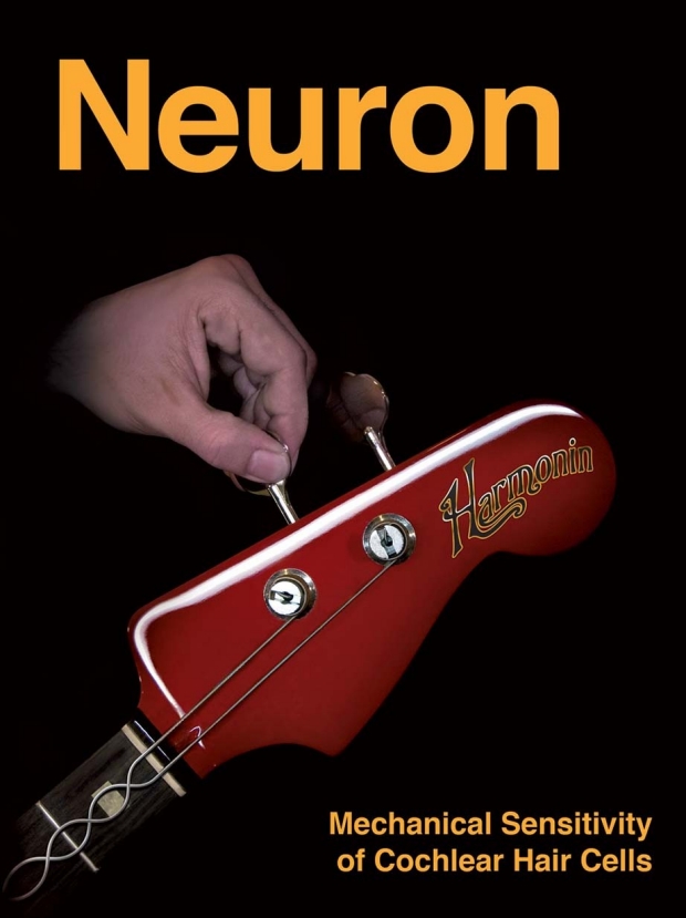
As in the strings of a guitar, tension must be adjusted in the mechanotransduction machinery of cochlear hair cells, which is essential for our ability to perceive sound. Tension in guitar strings is adjusted at the headstock in the upper part of the instrument. In hair cells, helical tip-link filaments, which gate mechanotransduction channels, terminate at their upper end in electron-dense structures. In the current issue (May 14, 2009), Grillet et al. (pp. 375–387) show that the PDZ-domain protein harmonin localizes to this structure and establishes tension in the hair cell's mechanotransduction machinery. Mutations in the gene for harmonin are linked to inherited forms of deafness, suggesting that defects in mechanotransduction are the underlying cause for some forms of the human disease. (Cover graphic design: johnforceps.com).

Mechanosensory hair cells of the cochlea play an essential role in converting mechanical vibrations into electrical signals that are subsequently transferred to afferent neurons. On pages 220–229 (April 2012, V35 issue 4), Piotr Kazmierczak and Ulrich Müller review recent studies that have provided insight into the molecular building-blocks of the mechanotransduction machinery. The cover design is a graphical representation of sound waves entering the cochlear of the inner ear. Image credit: Nicolas Grillet.