Research and Publications
Current Research & Scholarly Interests
Thyroid Eye Disease (Video: TEPEZZA® - FDA-Approved Treatment)
Adenoid Cystic Carcinoma of the Lacrimal Gland
Lacrimal Gland Stimulation for the Treatment of Dry Eyes
Neurostimulation
Orbital Tumors
Floppy Eyelid Syndrome and Obstructive Sleep Apnea
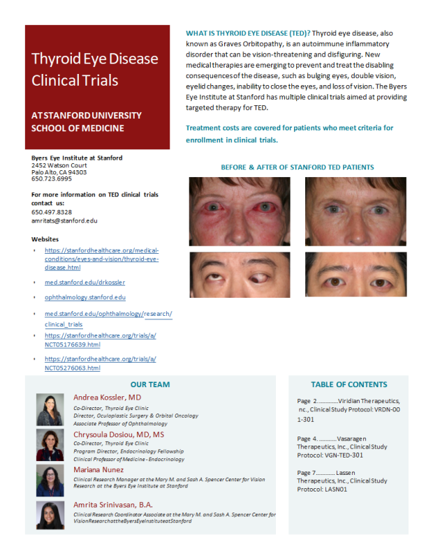
View more information about our Thyroid Eye Disease Clinical Trials in our pamphlet or in the trial details below.
1. A phase 3b/4 double masked, randomized clinical trials to evaluate the safety, efficacy and tolerability of different dosing duration of Teprotumumab
Status: Active
Research Coordinator: Farheen (fqshaikh@stanford.edu)
For eligibility and more information, please see NCT05002998.
Background: This is a double-masked, randomized, parallel-assignment, multicenter trial examining the safety and tolerability of teprotumumab in the treatment of Thyroid Eye Disease (TED) in adult participants. This international, Phase 3b/4 trial is being conducted to fulfill an FDA post-marketing requirement for a descriptive trial to evaluate the safety, efficacy and need for re-treatment of 3 different teprotumumab treatment durations for TED. In addition, serum samples from participants with a Baseline Clinical Activity Score (CAS) ≥3 will be evaluated for biomarkers of disease.
2. A Phase 2b, Randomized, Double-Mask, Placebo Controlled, Study to Evaluate the Safety, Pharmacokinetics and Efficacy of Oral Linsitinib in Subjects with Active, Moderate to Severe Thyroid Eye Disease
Status: Active
Co-PI: Chrysoula Dosiou, MD, MS
Research Coordinator: Farheen (fqshaikh@stanford.edu)
For eligibility and more information, please see NCT05276063.
Background: The overall objective is to study the safety, pharmacokinetics and efficacy of linsitinib (a small molecule IGF-1R inhibitor) administered orally twice daily (BID) vs. placebo, at 24 weeks in the treatment of subjects with active, moderate to severe thyroid eye disease (TED).
3. Evaluate the safety, tolerability and Efficacy of VRDN-001, a monoclonal antibody in patients with Thyroid Eye disease
Status: Active
Research Coordinator: Farheen (fqshaikh@stanford.edu)
For eligibility and more information, please see NCT05176639.
Background: The investigational drug, VRDN-001, is a monoclonal antibody that inhibits the activity of a cell surface receptor called insulin-like growth factor-1 receptor (IGF-1R). Inhibition of IGF-1R may help to reduce the inflammation and associated tissue swelling that occurs in patients with thyroid eye disease (TED). This clinical trial will evaluate the safety, tolerability and pharmacokinetics (the concentration of drug in the blood over time) of VRDN-001 in healthy volunteers and in patients with TED. Study participants with TED will also be evaluated over time for changes in their signs and symptoms of TED compared to their baseline measurements.
Surgical Idiopathic Intracranial Hypertension Treatment Trial
Randomized trial of adults (≥18 years old) with idiopathic intracranial hypertension and moderate to severe visual loss without substantial recent treatment who are randomly assigned to (1) medical therapy, (2) medical therapy plus ONSF, or (3) medical therapy plus VPS. The primary outcome is visual field mean deviation change at first of Month 6 (26 weeks) or time of treatment failure of the eligible eye(s), followed by a continuation study to assess time to treatment failure. The determination of eligible eye(s) is based on meeting the eligibility criteria at baseline.
Stanford is currently not accepting patients for this trial.
Stanford Investigator(s):
Intervention(s):
- drug: Acetazolamide
- procedure: Optic Nerve Sheath Fenestration
- procedure: Ventriculoperitoneal CSF Shunting
Eligibility
Inclusion Criteria:
- Subject Eligibility Criteria Inclusion Criteria
1. Diagnosis of IIH by modified Dandy criteria (Table 4)
2. Age 18 to <64 years at time of consent
3. Age 18 to <61 years at time of diagnosis (time of diagnosis is the time at which
the patient meets the modified Dandy criteria, usually after the lumbar puncture
results are reviewed)
4. Presence of bilateral papilledema
5. Lumbar puncture within 6 weeks of screening visit or completed as part of
screening: Opening CSF pressure >250 mmH2O or 200 to 250 mmH2O with at least one
of the following:
- Pulse synchronous tinnitus
- Cranial nerve VI palsy
- Echography for disc drusen negative and no other disc anomalies mimicking
disc edema present
- Magnetic Resonance Venography (MRV) with lateral sinus collapse/stenosis,
partially empty sella turcica on coronal or sagittal views of MRI, and optic
nerve sheaths with filled out CSF spaces next to the globe on T2 weighted
axial MRI scans If the patient was treated with intracranial pressure
lowering agents (e.g., acetazolamide) prior to obtaining a lumbar puncture,
the agent(s) must be discontinued for at least 24 hours prior to performing
the diagnostic lumbar puncture.
6. At least one eye meeting all eligible eye inclusion criteria and no exclusion
criteria.
7. Able to provide informed consent
8. Investigator believes participant is a good candidate for the study, including
the probability of returning for follow-up.
- Eye-Level Eligibility Criteria Subjects must have at least one eye meeting all of the
inclusion criteria and none of the exclusion criteria.
If both eyes meet eligibility criteria at the baseline examination, both will be included
in the primary outcome analysis.
Inclusion
1. Visual field loss meeting the following criteria based on two full threshold 24-2 size
V tests reviewed by the VFRC:
- PMD from -6 decibel (dB) to -27 dB
- Reproducible visual loss present on automated perimetry including no more than
15% false positive response
2. Visual acuity better than 20/200 (39 or more letters correct)
Exclusion Criteria:
- Subject Exclusion Criteria Exclusion Criteria
1. Treatment of IIH within the past 3 months with either (1) the maximally tolerated
dosage of acetazolamide for at least one week or (2) more than one month of
acetazolamide with a cumulative dosage of more than 45 grams 'Maximally-tolerated
dose' is defined as dosage was reached where dosage could not be increased
further either because of side effects or because a daily total dosage of 4 grams
per day was reached.
If individual discontinued acetazolamide in the past due to side effects,
individual is only eligible if investigator believes that the individual is
likely to tolerate acetazolamide, as it will be prescribed in the study.
2. Treatment of IIH within the past 3 months with either (1) the maximally tolerated
dosage of methazolamide for at least one week or (2) more than one month of
methazolamide with a cumulative dosage of more than 4.5 grams
'Maximally-tolerated dose' is defined as dosage was reached where dosage could
not be increased further either because of side effects or because a daily total
dosage of 400 mg per day was reached.
3. Treatment with topiramate within two months and average cumulative dosage for the
preceding month of more than 700 mg per week
4. Previous surgery for IIH, including ONSF, CSF shunting, subtemporal
decompression, or venous sinus stenting; gastric surgery for obesity is allowed
5. Abnormalities on neurologic examination except for papilledema and its related
visual loss or cranial nerve VI to VII paresis; if other abnormalities are
present, the patient will need to be discussed with the Study Director (SD) for
study entry.
6. Abnormal CT or MRI scan (intracranial mass, hydrocephalus, dural sinus thrombus,
or arteriovenous malformation) other than findings known to occur with increased
intracranial pressure. Abnormalities on MRI that are not known to cause increased
intracranial pressure are acceptable.
7. Abnormal CSF contents: increased cells: > 8 cells; elevated protein: > 45 mg%;
low glucose: < 30 mg% (If the lumbar puncture produces a cell count compatible
with a traumatic needle insertion, the patient does not need to be excluded if
the CSF white blood cell count (WBC) after correction is 8 cells/mm3 or less -
see Manual of Procedures (MOP) for calculation. If > 8 cells or > 45mg% in CSF
protein are documented in the CSF or calculated after conversion from a traumatic
lumbar puncture, the patient can be discussed with the Study Director for
possible inclusion.)
8. Abnormal blood work-up indicating a medical or systemic condition associated with
raised intracranial pressure
9. Diabetes mellitus with diabetic retinopathy
10. Ingestion of a drug or substance, or presence of a disorder, that has been
associated with increased intracranial pressure within 2 months of diagnosis,
such as lithium, vitamin A related products (e.g., Retin-A), or various cyclines
(see MOP for conditions and drugs)
11. Laboratory test results showing severe anemia, leukopenia or thrombocytopenia,
renal failure, or hepatic disease, based on the Site Investigator's judgment
12. Other condition requiring continued use of oral, I.V. or injectable steroids
(nasal, inhaled, or topical steroids are allowed since the systemic effects are
small). Patients with a condition that resulted in recent or current use of
steroids but may be safely tapered off will be handled on a case-by-case basis
after discussion with Study Director/co-Director. See Manual of Procedures (MOP)
for details.
13. Presence of a medical condition that would contraindicate use of acetazolamide or
furosemide or significantly increase surgical risk
14. Pregnancy or unwillingness for a subject of childbearing potential to use
contraception during the first 6 months of the study Women of childbearing
potential must use an acceptable form of birth control during the first 6 months
of the study. Acceptable forms include oral contraceptives, transdermal
contraceptives, diaphragm, intrauterine devices (IUDs), condoms with spermicide,
documented surgical sterilization of either the subject or their partner, or
abstinence.
15. Presence of a physical, mental, or social condition likely to affect follow-up
(drug addiction, terminal illness, no telephone, homeless)
16. Anticipation of a move from the site area within six months and unwillingness to
return for follow-up at a SIGHT study site
17. Allergy to pupil dilating drops or narrow angles precluding safe dilation
18. Presence of a condition that contraindicates general anesthesia
19. Participation in an investigational trial within 30 days of enrollment that
involved treatment with any systemic drug therapy or therapy that affects the
eligible eye(s)
- Eye Level Exclusion Criteria Exclusion
1. Intraocular pressure currently >28 mm Hg or >30 mm Hg at any time in the past
2. Refractive error of more than -6.00 or more than +6.00 sphere or more than 3.00
cylinder with the following exceptions:
- Eyes with more than 6.00 D of myopia but less than 8.00 D of myopia are
eligible if: 1) there are no abnormalities on ophthalmoscopy related to
myopia that are associated with visual loss (such as staphyloma, retinal
thinning in the posterior pole, or more than mild optic disc tilt), and 2)
the individual will wear a contact lens for all perimetry examinations with
the appropriate correction.
- Eyes with more than 6.00 D of hyperopia but less than 8.00 D of hyperopia
are eligible if: 1) there is an unambiguous characteristic halo of
peripapillary edema as opposed to features of a small crowded disc or other
hyperopic change related to visual loss determined by the Site Investigator
or the Photographic Reading Center (PRC) Director (or his designate), and 2)
the individual will wear a contact lens for all perimetry examinations with
the appropriate correction (which can be corrected for perimetry or with the
patient's own contact lens with over correction by lens at the perimeter).
Note: Refractive error exclusion and exceptions refer to sphere not spherical
equivalent, with cylinder expressed in plus format.
3. Other disorders causing visual loss except for refractive error and amblyopia,
including cells in the vitreous or iritis
4. Large optic disc drusen on exam or known in previous history (small drusen of the
disc can occur with longstanding papilledema and are allowed if not so numerous
that investigator determines they are contributing to vision loss)
Ages Eligible for Study
18 Years - 63 Years
Genders Eligible for Study
All
Not currently accepting new patients for this trial
Contact Information
Stanford University
School of Medicine
300 Pasteur Drive
Stanford,
CA
94305
Mariana Nunez
650-497-7846
Not Recruiting
Publications
-
An Analysis of Solicitations from Predatory Journals in Ophthalmology.
American journal of ophthalmology
Justin, G. A., Huang, C., Nguyen, M. K., Lee, J., Seddon, I., Wesley, T. A., Bakri, S. J., Campbell, J. P., Cavuoto, K., Collins, M., Gedde, S. J., Kossler, A. L., Milman, T., Shukla, A., Sridhar, J., Syed, Z. A., Williams, B. K., Woreta, F. A., Patel, S. N., Yonekawa, Y.
2024
Hide
More
Abstract
To evaluate trends associated with email communication from potentially predatory publishers to faculty in ophthalmology.Cross sectional study METHODS: Ophthalmologists (n=14) from various subspecialties and institutions were recruited to participate. Participants identified unsolicited emails they had received originating from publishers in May 2021. Information collected included details on email contents and publisher organizations. Trends in communications from predatory publishers were evaluated.Over a 30-day study period, a total of 1813 emails were received from 383 unique publishers and 696 unique journals with a mean (SD) of 4.73 (2.46) emails received per day per participant. Of the 1813 emails identified, 242 (13%) emails were invitations to conferences, whereas 1440 (80%) were solicitations for article submissions to open-access pay-to-publish journals. A total of 522 (29.0%) emails were related to ophthalmology, and reference to a prior publication of the participant occurred in 262 emails (14%). Of the 696 unique journals identified, 174 (25%) journals were indexed on PubMed and 426 (61%) were listed on Beall's list. When comparing journals listed on PubMed versus those that were not, PubMed indexed journals had a higher impact factor (2.1 vs 1.5, p=0.002), were less likely to use "greetings" (76% vs 91%, p<0.001), had fewer spelling/grammar errors (40% vs 51%, p=0.01), and were less likely to offer rapid publication (16% vs 25%, p=0.02).Unsolicited requests to publish occur frequently and may diminish the quality of the scientific literature. We encourage individuals in ophthalmology to be aware of these trends in predatory publishing.
View details for DOI 10.1016/j.ajo.2024.02.030
View details for PubMedID 38490339
-
Association of ocular manifestations of Marfan syndrome with cardiovascular complications.
American journal of ophthalmology
Tran, E. M., Wai, K. M., Kossler, A. L., Mruthyunjaya, P., Rahimy, E., Koo, E. B.
2024
Hide
More
Abstract
To evaluate associations between ocular manifestations of Marfan syndrome and cardiovascular complications.retrospective cohort study METHODS: Setting: TriNetX Analytics platform, a federated health research network of aggregated deidentified electronic health record data of over 119 million patients.Patients diagnosed with Marfan syndrome.Univariate logistic regression models were used to evaluate the association of ocular manifestations of Marfan syndrome (such as retinal tears/detachment, lens dislocation, and myopia), with cardiovascular comorbidities. Additional sensitivity analyses were performed using propensity matching.Odds ratio and 95% confidence intervals for incidence of cardiovascular comorbidities (including aortic dissection, valvular disease, and arrhythmias) following diagnosis of Marfan syndrome.19,105 patients were identified that were diagnosed with Marfan disease without ocular manifestations, and an additional 3,887 Marfan patients with ocular comorbidities. Patients who were diagnosed with ocular disease included 883 with ectopic lens, 417 with retinal tear or detachment, 683 with aphakia, 534 with pseudophakia, and 2,465 with myopia. Patients with any ocular manifestations of Marfan were significantly more likely to be diagnosed with all cardiovascular comorbidities modeled including aortic aneurysm and dissection (OR=2.035; p=<0.0001), mitral valve prolapse (OR=2.725; p= p=<0.0001), tricuspid valve disorders (OR=2.142; p=<0.0001), cardiac arrhythmias (OR=1.836; p=<0.0001), and all cardiovascular outcomes combined (OR=2.194; p=<0.0001).In a large and diverse cohort of patients with Marfan syndrome, ocular manifestations of the disorder appear strongly associated with cardiovascular comorbidities.
View details for DOI 10.1016/j.ajo.2024.02.023
View details for PubMedID 38403098
-
Reply.
Ophthalmology
Shah, S. A., Amarikwa, L., Sears, C. M., Clauss, K. D., Rajjoub, R. D., Kang, J. Y., Tamhankar, M. A., Briceño, C. A., Harrison, A. R., Cockerham, K. P., Wester, S. T., Douglas, R. S., Dosiou, C., Kossler, A. L.
2024
Hide
More
View details for DOI 10.1016/j.ophtha.2024.01.003
View details for PubMedID 38349298
-
Anatomic and Surgical Considerations in the Management of a Sellar and Suprasellar Arachnoid Cyst: 2-Dimensional Operative Video.
Operative neurosurgery (Hagerstown, Md.)
Rychen, J., Constanzo, F., Chan, D., Kossler, A. L., Fernandez-Miranda, J. C.
2024
Hide
More
View details for DOI 10.1227/ons.0000000000001061
View details for PubMedID 38198191
-
Teprotumumab for the Treatment of Recalcitrant Thyroid Eye Disease.
Ophthalmic plastic and reconstructive surgery
Men, C. J., Amarikwa, L., Pham, B., Sears, C., Clauss, K., Lee, B. W., Lee, W. W., Pasol, J., Ugradar, S., Shinder, R., Cockerham, K., Wester, S., Douglas, R., Kossler, A. L.
2023
Hide
More
Abstract
Teprotumumab, an insulin-like growth factor 1 receptor monoclonal antibody, is FDA-approved to treat thyroid eye disease (TED). The initial clinical trials excluded patients with previous orbital irradiation, surgery, glucocorticoid use (cumulative dose >1 gm), or prior biologic treatment. Information on the use of teprotumumab for patients who failed prior therapy is limited. Our purpose is to characterize the efficacy of teprotumumab for the treatment of recalcitrant TED.This is a multicenter retrospective study of all patients treated with teprotumumab for moderate-to-severe TED after failing conventional therapy with corticosteroids, orbital radiation, surgical decompression, biologics, or other steroid-sparing medications. Treatment failure was defined as an incomplete response to or reactivation after previous treatment. Only patients who received at least 4 infusions of teprotumumab were included in the analysis. Primary outcome measures comprised proptosis response (≥2 mm reduction in the study eye without a similar increase in the other eye), clinical activity score (CAS) response (≥2-point reduction in CAS), and diplopia response (≥1 point improvement in Gorman diplopia score in patients with baseline diplopia) following treatment. Adverse events and risk factors for recalcitrant disease were also evaluated.Sixty-six patients were included in this study, 46 females and 20 males. Average age was 59.3 years (range 29-93). The mean duration of disease from TED diagnosis to first infusion was 57.8 months. The proptosis, CAS, and diplopia responses in this recalcitrant patient population were 85.9%, 93.8%, and 69.1%, respectively. Patients experienced a mean reduction in proptosis of 3.1 ± 2.4 mm and a mean improvement in CAS of 3.8 ± 1.6. Patients who underwent prior decompression surgery experienced a statistically significant decrease in diplopia response (46.7% vs. 77.5%, p = 0.014) and proptosis response (75.0% vs. 90.9%, p = 0.045) when compared with nondecompression patients. Additionally, there were no significant differences in proptosis, CAS, and diplopia responses between patients with acute (defined as disease duration <1 year) versus chronic (disease duration ≥1 year) TED. While most adverse events were mild to moderate, 4 patients reported serious adverse events related to persistent hearing loss.Patients with recalcitrant TED demonstrated a significant improvement after teprotumumab in each of the primary study outcomes. The degree of proptosis reduction, diplopia response, and CAS improvement in the recalcitrant group were similar to those of treatment-naïve patients from the pivotal clinical trials. Patients with a prior history of orbital decompression, however, demonstrated poor improvement in diplopia and less reduction in proptosis than surgery naïve patients. These results indicate that teprotumumab is a treatment option for the treatment of patients with TED recalcitrant to prior medical therapies.
View details for DOI 10.1097/IOP.0000000000002564
View details for PubMedID 37972960
-
Teprotumumab-Related Adverse Events in Thyroid Eye Disease: A Multi-Center Study.
Ophthalmology
Shah, S. A., Amarikwa, L., Sears, C. M., Clauss, K. D., Rajjoub, R. D., Kang, J. Y., Tamhankar, M. A., Briceño, C. A., Harrison, A. R., Dosiou, C. C., Cockerham, K. P., Wester, S. T., Douglas, R. S., Kossler, A. L.
2023
Hide
More
Abstract
To assess the duration, incidence, reversibility, and severity of adverse events (AEs) in patients with thyroid eye disease (TED) treated with teprotumumab.Multi-center retrospective observational cohort study.Patients with TED of all stages and activity levels treated with at least 4 infusions of teprotumumab.Patients were treated with teprotumumab between February 2020 and October 2022 at 6 tertiary centers. AE metrics were solicited and recorded at each visit. AEs were grouped according to the United States FDA Adverse Event Reporting System.Primary outcomes measure: AE incidence and onset.AE severity, reversibility, duration, proptosis response, clinical activity score (CAS) reduction, and Gorman diplopia score (GDS) improvement.The study evaluated 131 patients. Proptosis improved by 2mm or more in 77% (101/131) of patients with 3.0±2.1mm average proptosis improvement and 3.2 points average CAS reduction. GDS improved by at least one point for 50% (36/72) of patients with baseline diplopia. AEs occurred in 81.7% (107/131) of patients. Patients had a median of 4 AEs. Most patients' AEs were mild (74.0%, 97/131), 28.2% (37/131) moderate, and 8.4% (11/131) severe. Mean interval AE onset was 7.9 weeks after the first infusion. Resolved AEs had a mean duration of 17.6 weeks. Forty-six percent (60/131) of patients had at least 1 persistent AE at last follow-up. Patients had a mean follow-up of 70.2±38.5 weeks after the first infusion. The most common type of AEs was musculoskeletal (58.0%, 76/131), followed by gastrointestinal (38.2%, 50/131), skin (38.2%, 50/131), ear and labyrinth (30.5%, 40/131), nervous system (20.6%, 27/131), metabolic (15.3%, 20/131), and reproductive system (12.2%, 16/131). Sixteen patients (12.2%) discontinued therapy due to AEs, including hearing loss (n=4), inflammatory bowel disease flare (n=2), hyperglycemia (n=1), muscle spasms (n=1), and multiple AEs (n=8).AEs are commonly reported while receiving teprotumumab treatment. Most are mild and reversible; however, serious AEs can occur and may warrant treatment cessation. Treating physicians should inform patients about the AE risk, properly screen patients prior to treatment, monitor patients closely throughout therapy, and understand how to manage AEs should they develop.
View details for DOI 10.1016/j.ophtha.2023.10.018
View details for PubMedID 37852417
-
Automated Detection of Dysthyroid Optic Neuropathy in Graves' Ophthalmopathy with Computed Tomography (CT) Scans by Convolutional Neural Networks
Hung, J., Luo, A., Deng, Y., Chung, C., Fuh, C., Perera, C., Myung, D., Kossler, A., Liao, S.
ASSOC RESEARCH VISION OPHTHALMOLOGY INC. 2023
Hide
More
View details for Web of Science ID 001053758307020
-
Characteristics of Predatory Publishing Solicitation in Ophthalmology
Nguyen, M., Huang, C., Lee, J., Seddon, I., Bakri, S., Syed, Z., Woreta, F., Shukla, A., Cavuoto, K., Collins, M., Williams, B., Kossler, A., Campbell, J., Patel, S., Justin, G., Yonekawa, Y.
ASSOC RESEARCH VISION OPHTHALMOLOGY INC. 2023
Hide
More
View details for Web of Science ID 001053795607128
-
Accuracy of Digital Image Analysis for Diagnosing IgG4 Related Ophthalmic Disease
Charoenkijkajorn, C., Gill, H., Glory, B., Shi, W., Homer, N., Men, C., Kossler, A., Wu, A., Lin, J.
ASSOC RESEARCH VISION OPHTHALMOLOGY INC. 2023
Hide
More
View details for Web of Science ID 001053795606146
-
The use of Google Trends and Twitter data as a tool for evaluating public interest in hyaluronic acid eyelid filler.
Journal of clinical and translational research
Cohen, S. A., Kossler, A. L.
2023; 9 (2): 76-83
Hide
More
Abstract
Google Trends and the Twitter Academic Research Product Tract (TARPT) are free, online tools that can be used to evaluate public interest in plastic surgery procedures.The aim of the study was to evaluate the correlation between online public interest in hyaluronic acid eyelid filler on two popular web platforms (Google and Twitter) and hyaluronic acid filler procedure volumes in the United States.The Google Trends database and the TARPT tool were used to calculate the number of annual Google searches and Twitter tweets, respectively, related to 10 search terms associated with hyaluronic acid eyelid filler injections from January 2010 to December 2020. Annual procedure volumes for hyaluronic acid filler injections were obtained from the American Society of Plastic Surgery (ASPS). Univariate linear regression was used to correlate Google searches to ASPS procedure volumes and Twitter tweet volumes to ASPS procedure volumes.Significant positive correlations were found between Google Trends data and ASPS procedure volumes for 8/10 search terms and between Twitter tweet volumes and ASPS procedure volumes for 6/10 search terms, respectively. Online public interest in eyelid filler related search terms increased significantly over time according to an exponential model (P < 0.0001).We observed statistically significant positive associations between public interest related to eyelid filler on two online platforms, Google and Twitter, and hyaluronic acid soft-tissue filler procedure volumes. The Google Trends and TARPT databases represent free information sources for surgeons that may be used to inform marketing and advertising decisions and to anticipate patient inquiries during the patient encounter.Information provided by the Google Trends and TARPT tools can be used by surgeons to (1) inform marketing and advertising strategies and (2) gain insight into which procedures patients are researching during a given time period, preparing them to best address the evolving needs of patients.
View details for PubMedID 37034000
View details for PubMedCentralID PMC10075089