A Subset of Cells That Can Break Your Heart
By Adrienne Mueller, PhD
September 11, 2020
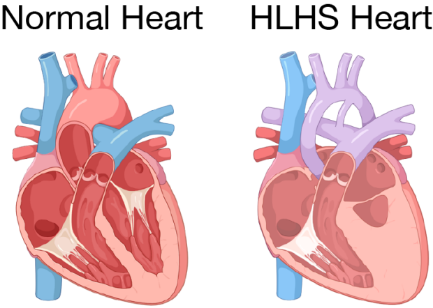
Hypoplastic left heart syndrome (HLHS) is characterized by the underdevelopment of heart valves as well as the left ventricle, the main pumping chamber of the heart.
Every year, approximately 1 out of every 3850 babies in the US is born with a heart defect called hypoplastic left heart syndrome (HLHS). This disorder is characterized by the underdevelopment of heart valves as well as the left ventricle, the main pumping chamber of the heart (Figure). However, the cause for why these hearts develop abnormally is not well understood. Scientists have long sought the mechanism responsible for the developmental abnormalities in the heart muscle of HLHS patients, however, the cells that exhibit deficits that could help explain the malformation seen in HLHS hearts remain elusive. Recently, a group of Stanford Cardiovascular Institute and affiliated researchers report the identification of endocardial cells as a key cell type in heart of HLHS patients responsible for the onset of this disease.
Endocardial cells are specialized cells that form the innermost layer of the heart wall, also known as the endocardium. The endocardium is very important for heart development during which the endocardium facilitates the formation and maturation of structural parts of the heart like the heart valves, the septum that divides the left and right-side of the heart chambers, and the coronary arteries. Interactions between endocardial cells and heart muscle cells are also important for maturation of the heart chambers. Genetic ablation of endocardial cells results in ventricular malformation. Since HLHS is characterized by structural abnormalities in the valves and under-development of the left ventricle, it has been speculated that a defect in the endocardium may be the mechanism underlying HLHS.
To test this hypothesis, a group of scientists, led by co-first authors Yifei Miao, PhD and Lei Tian, PhD and senior author Mingxia Gu, MD, PhD, compared the endocardial cells derived from human induced pluripotent stem cell (hiPSCs) from healthy individuals and individuals with HLHS. In collaboration from members of Sean M. Wu, MD, PhD lab, the investigative team sought to identify differences in the genes expressed between these two groups. The investigators employed single-cell RNA-sequencing to profile gene expressed in endocardial cells derived from HLHS patients and normally-developing endocardial cells. By carefully profiling the expression of the different cells they collected, the investigators identified a subpopulation of endocardial cells that were developmentally impaired in HLHS hiPSC-derived endocardial cells. These investigators then went on to demonstrate that the HLHS hiPSC-derived endocardial cells were functionally impaired in several ways. First, several key signaling pathways important for valve development were suppressed. Second, the interface between the endocardium and the heart muscle was abnormal. And, third, the ability to develop new vessels was impaired. Additionally, this abnormal population of hiPSC-derived HLHS endocardial cells had a significant impact on heart muscle cell development: when young heart muscle cells were cultured next to hiPSC-derived HLHS endocardial cells, fewer heart muscle cells grew and those that did grow did not properly mature.
Miao and Tian et al’s study, recently reported in Cell Stem Cell, has uncovered a mechanism that helps to explain the congenital heart defects seen in HLHS patients (i.e. the presence of faulty endocardial cells causes developmental impairment in the endocardium). Not only does this study help explain the cause of HLHS, but it suggests that approaches that focus on endocardial function could be a fruitful avenue for treating other heart diseases.
Other Stanford Cardiovascular Institute-affiliated authors include Marcy Martin, Sharon L. Paige, Francisco X. Galdos, Alyssa Klein, Hao Zhang, Ning Ma, Soah Lee, Jan-Renier Moonen, Joseph C. Wu, and Marlene Rabinovitch.

Dr. Yifei Miao
Dr. Lei Tian

Dr. Mingxia Gu