ISMRM Honors 2019
Young Investigator Award
Akshay Chaudhari
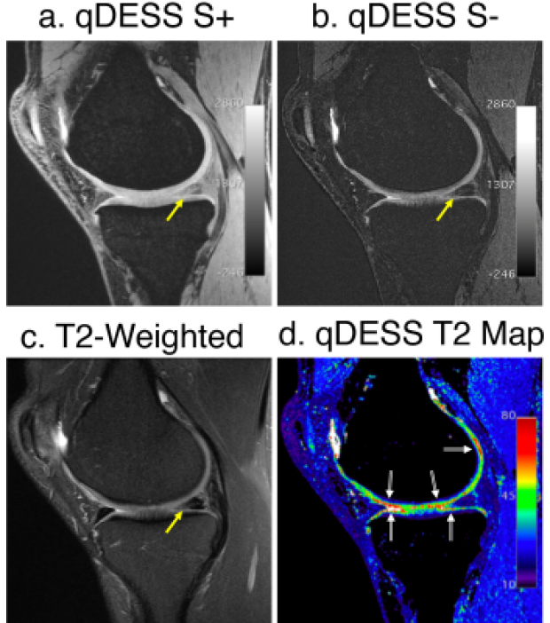
Akshay Chaudhari, Zhongnan Fang, Murray Grissom, Bragi Sveinsson, Jeff Wood, Christopher Beaulieu, Edwin Oei, Jarrett Rosenberg, Feliks Kogan, Jin Hyung Lee, Marcus Alley, Garry Gold, Kathryn Stevens, Brian Hargreaves
Most knee MRI protocols require 20+ minutes of scan time, leading to interest in expedited protocols. Here, we first demonstrate in a study with 35 patients and 5 readers that for diagnostic knee MRI, a 3D 5-minute quantitative double-echo steady-state (qDESS) sequence has high agreement with the conventional sequences, where the addition of a proton-density-weighted sequence engenders near-perfect agreement. In a second study with 51 patients and 2 readers, we demonstrate that qDESS with two-fold enhanced slice resolution using deep-learning-based super-resolution and T2 maps has high agreement with the conventional sequences, where both methods have similar agreement with arthroscopic findings.
Summa Cum Laude

Extreme MRI: Super-High-Res Dynamic Volumetric MRI from Continuous Non-Gated Acquisition
Frank Ong, Xucheng Zhu, Peder Larson, Joseph Cheng, Shreyas Vasanawala, Michael Lustig
The goal of this work is to recover transient dynamics in 3D dynamic MRI by reconstructing images with near-millimeter spatial resolution and sub-second temporal resolution without gating. This setting poses two major challenges: extreme undersampling and extreme computational/memory cost. To achieve this “extreme MRI”, we propose two innovations: explicit multi-scale low rank matrix factorization to regularize the problem and reduce memory usage, and stochastic optimization to reduce computation. We demonstrate the feasibility of the proposed method in DCE imaging acquired with 3D cones trajectory and lung imaging acquired with 3D UTE radial trajectory.
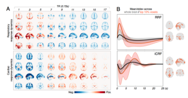
Resting-State “Physiological” Networks
Jingyuan Chen, Laura Lewis, Catie Chang, Nina Fultz, Ned Ohringer, Bruce Rosen, Jonathan Polimeni
In this abstract, we offer a systemic characterization of the spatiotemporal patterns of fMRI signals subsequent to slow fluctuations in respiratory volume and heart rate. We show that these slow physiological dynamics contain structured network patterns that are somewhat consistent across individuals. We also show that global signal regression (GSR) may introduce anti-correlating patterns of the physiological dynamics to the final observations of functional connectivity.
Magna Cum Laude

GagCEST at 3T Can Detect Cartilage Differences Between Healthy and OA Subjects
Elka Rubin, Lauren Watkins, Valentina Mazzoli, Arjun Desai, Gabe Ho, Feliks Kogan, Scott Ulrich, Julie Kolesar, Scott Delp, Gary Beaupre, Garry Gold
Chemical exchange saturation transfer of GAG (gagCEST) is a quantitative MR technique that is a useful biomarker for assessing GAG content at 7T. However, its utility at 3T remains unclear. In this study, we compare gagCEST asymmetry values of healthy and osteoarthritic subjects scanned at 3T. Comparisons between healthy and OA subjects indicate a significant difference in the average gagCEST signal across the medial and lateral anterior and medial weight-bearing regions of the femoral cartilage. The results of this study suggest that there is potential for use of gagCEST in the study of OA at 3T.

Qiyuan Tian, Chanon Ngamsombat, Berkin Bilgic, Qiuyun Fan, Yuxin Hu, Jennifer McNab, Thomas Witzel, Kawin Setsompop, Jonathan Polimeni, Susie Huang
Tremor suppression in the hands of patients with essential tremor can be achieved by lesioning the ventral intermediate nucleus (Vim) of the thalamus using transcranial MR-guided focused ultrasound. Recent work has shown that diffusion MR tractography identifies the Vim more precisely and predicts the degree of tremor suppression. Here, we trained a convolutional neural network (CNN) to automatically segment relevant regions-of-interest including the thalamus, red nucleus, dentate nucleus and handknob region for probabilistic tractography to identify the Vim. We applied the CNN to 200 HCP healthy subjects and created a tractography-based atlas of Vim location, which could aid in neurosurgical guidance.
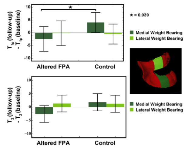
Gait retraining as a conservative treatment for medial knee OA: preliminary findings
Valentina Mazzoli, Scott Uhlrich, Elka Rubin, Feliks Kogan, Brian Heargraves, Scott Delp, Gary Beaupre, Garry Gold
Osteoarthritis is a major societal burden and is associated with pain and disability. To cope with the “osteoarthritis epidemic” and its high associated costs, there is a need for new conservative treatments. This study investigates the potential of gait retraining with altered foot progression angle as one inexpensive conservative treatment for medial knee osteoarthritis. Our results show that this treatment may be effective in reducing pain and improving the MRI outcomes in osteoarthritis patients. This suggests the potential of personalized gait retraining with altered foot progression angle as an inexpensive and effective conservative method for the management of osteoarthritis patients.

Five-minute single sequence comprehensive 4D pediatric ankle MRI with T2 Shuffling
Jonathan Tamir, Fida Wishah, Jesse Sandberg, Marcus Alley, Michael Lustig, Shreyas Vasanawala
The use of volumetric acquisitions for musculoskeletal MR in clinical settings has been limited due to blurring artifacts from T2 decay. T2 Shuffling (T2Sh), a redesigned 3D fast spin-echo technique that mitigates blurring, has been successfully applied to pediatric knee MRI in a clinical setting and used to streamline the pediatric knee exam. This work assesses a shortened T2Sh scan for pediatric ankle MRI with total scan time under 5 minutes. Our results show that T2Sh has the potential to provide a comprehensive diagnostic protocol in place of the conventional long 2D exam.
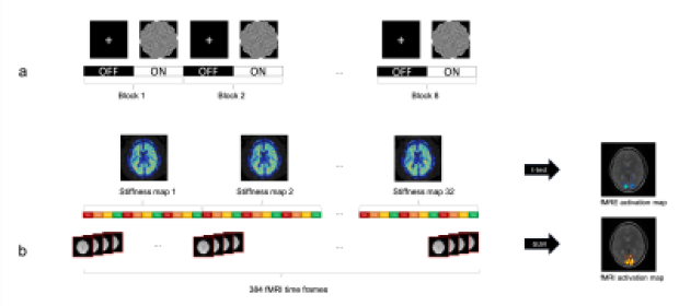
Patricia Lan, Kevin Glaser, Richard Ehman, Gary Glover
Here, we demonstrate a novel multi-modal method to simultaneously acquire robust fMRE and fMRI activation maps. A block paradigm of 24s ON (flashing checkerboard at 10Hz) and 24s OFF (fixation cross) was used and images were acquired with a single-shot spin-echo EPI MRE sequence. Our results show that tissue stiffness within the visual cortex increases 6-12% with visual stimuli. Furthermore, the fMRE and fMRI activation maps agree and overlap spatially within the visual cortex, providing convincing evidence that fMRE is possible in the cortex.
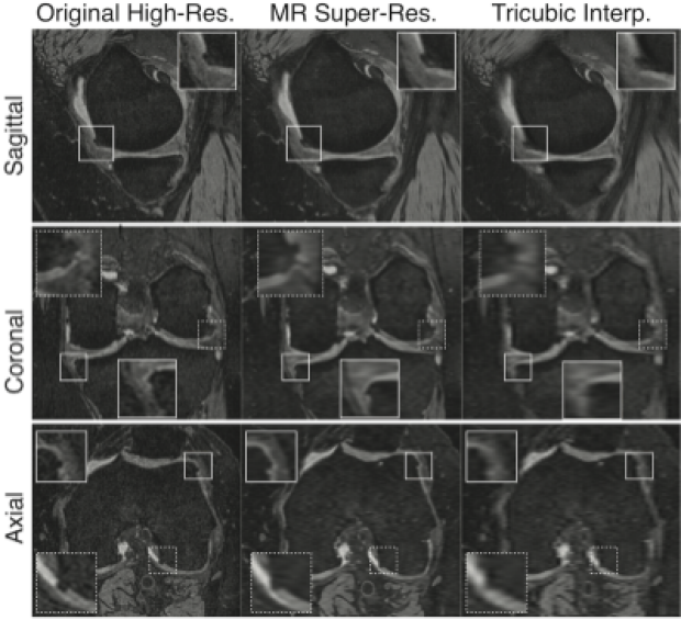
Evaluating the Use of Deep-Learning Super-Resolution for Obtaining Osteoarthritis Biomarkers
Akshay Chaudhari, Jeff Wood, Kathryn Stevens, Zhongnan Fang, Jin Lee, Garry Gold, Brian Hargreaves
The use high-resolution magnetic resolution imaging (MRI) is beneficial for acquiring quantitative biomarkers corresponding to osteoarthritis (OA) severity and progression. However, the long scan times of high-resolution sequences, such as double-echo steady-state (DESS) that was included in the Osteoarthritis Initiative, precludes their widespread adoption. Deep-learning-based super-resolution has the potential to transform low-resolution MRI that can be acquired faster, into high-resolution images. Using qualitative cartilage image quality, and quantitative cartilage morphometry and osteophyte detection, we have shown that deep-learning-based super-resolution can enhance DESS slice-resolution threefold and offer the same utility as the original high-resolution acquisition for obtaining OA biomarkers.

Functional Magnetic Resonance Electrical Impedance Tomography of Aplysia abdominal ganglion
Fanrui Fu, Munish Chauhan, Rosalind Sadleir
Functional Magnetic resonance electrical impedance tomography (fMREIT) has the potential to directly image neural activity. In our tests, we used the Aplysia abdominal ganglion (AAG) as a neuronal activity source. Potassium chloride (KCl) was used to modulate neuronal activity. Analysis of magnitude images, subtracted MREIT phase images and Laplacian of MREIT Bz images was performed to evaluate the difference in MREIT images with and without neuronal activity.
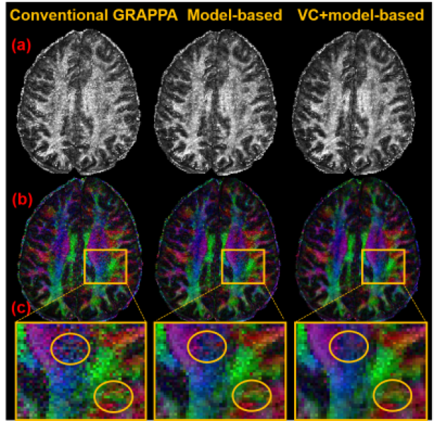
Simin Liu, Erpeng Dai, Zijing Dong, Hua Guo
Recently, generalized SLIce Dithered Enhanced Resolution (gSlider) has been proposed as a new acquisition strategy for isotropic high-resolution DTI. In this study, to further boost the SNR performance and reconstruction accuracy, the model-based DTI reconstruction method was merged into the gSlider reconstruction procedure. Moreover, virtual coil (VC) concept was also integrated into the proposed method to further improve the SNR of gSlider reconstruction. Compared with conventional GRAPPA, the superiority of the model-based method has been demonstrated by in vivo results, especially when in combination with the virtual coil concept.
Anita Jwa, Jonathan Goodman, Gary Glover
Transcranial direct current stimulation (tDCS) is gaining momentum both in the research community and in the general public as an attractive tool for neuromodulation. However, the mechanism of tDCS in the brain is not fully understood. In this study, we conducted a concurrent tDCS–magnetic resonance current density imaging (MRCDI) experiment to measure the primary direction and magnitude of current in a human brain undergoing direct current stimulation. Our results show that current flow deviates significantly from its desired distribution based on human head models and that the current mostly flows through the white matter and cerebrospinal fluid.

Reconstruction of multi-shot diffusion-weighted MRI using unrolled network with U-nets as priors
Yuxin Hu, Xinwei Shi, Qiyuan Tian, Hengkai Guo, Minda Deng, Miao Yu, Catherine Moran, Grant Yang, Jennifer McNab, Bruce Daniel, Brian Hargreaves
In this work, we demonstrated the feasibility of using deep neural networks for rapid multi-shot DWI reconstruction. An unrolled network with six U-nets, which operated in frequency and image domains alternatively, was shown to have the capability to remove aliasing artifacts from shot-to-shot phase variations, and achieved about 60-fold speed up and around 1% amplitude difference compared with conventional iterative reconstruction methods.
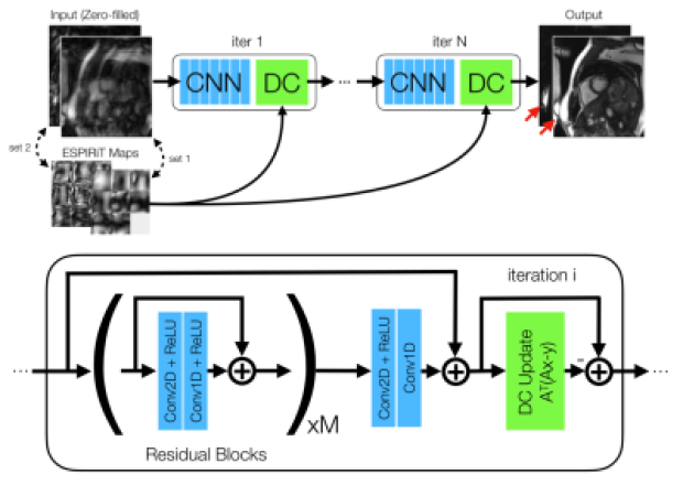
DL-ESPIRiT: Improving robustness to SENSE model errors in deep learning-based reconstruction
Christopher Sandino, Peng Lai, Shreyas Vasanawala, Joseph Cheng
Parallel imaging concepts, such as sensitivity encoding (SENSE), have been incorporated into DL reconstruction frameworks by augmenting the acquisition model with knowledge of the coil sensitivities. However, SENSE-based methods rely on accurate estimation of sensitivity maps; otherwise, residual aliasing may arise due to model errors such as in reduced field-of-view imaging. Here we propose DL-ESPIRiT, an ESPIRiT-based neural network architecture with improved robustness to model errors. We show that DL-ESPIRiT can reconstruct 10X accelerated 2D cardiac CINE data with higher fidelity and allow for more accurate automatic assessment of cardiovascular function than l1-ESPIRiT.

Post-treatment diffusion weighted imaging predicts ablation zone margins following MRI-guided high intensity focused ultrasound of the prostate.
Ryan Brunsing, Signy Holmes, Rachelle Bitton, Bruce Daniel, Geoffrey Sonn, Pejman Ghanouni
The efficacy and safety of magnetic resonance imaging-guided high intensity focused ultrasound (MRg-HIFU) treatment of intermediate risk prostate cancer is being assessed as part of a clinical trial. Non-perfused volume (NPV) on post-treatment contrast-enhanced imaging defines the zone of ablation, however contrast administration precludes further treatment due to concern for gadolinium dissociation. Thus, pre-contrast imaging tools which can predict NPV are of value. From a cohort of 19 men who underwent MFg-HIFU for treatment of prostate cancer, we show that post-treatment DWI can predict NPV, with the potential to increase confidence in predicting the ablation zone prior to contrast administration

Unsupervised Deep Basis Pursuit: Learning Reconstruction without Ground-Truth Data
Jonathan Tamir, Stella Yu, Michael Lustig
Basis pursuit is a compressed sensing optimization in which the l1-norm is minimized subject to model error constraints. Here we use a deep neural network prior instead of l1-regularization. Using known noise statistics, we jointly learn the prior and reconstruct images without access to ground-truth data. During training, we use alternating minimization across an unrolled iterative network and jointly solve for the neural network weights and training set image reconstructions. At inference, we fix the weights and pass the measurements through the network. We compare reconstruction performance between unsupervised and supervised (i.e. with ground-truth) methods. We hypothesize this technique could be used to learn reconstruction when ground-truth data are unavailable, such as in high-resolution dynamic MRI.
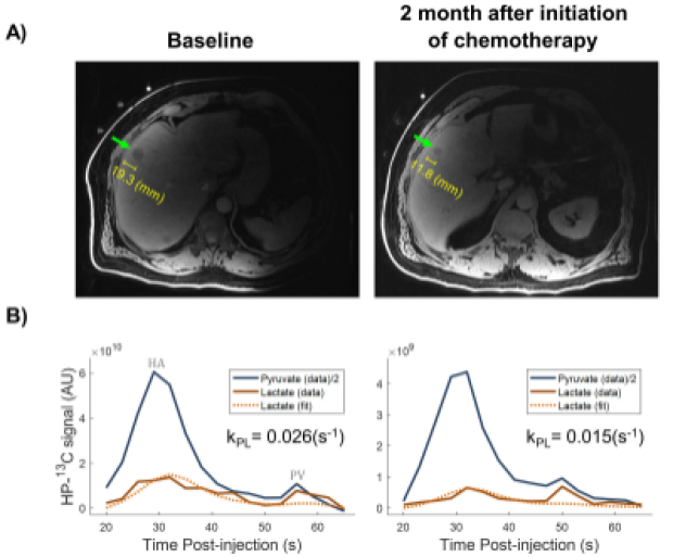
Hyperpolarized 13C MRI of Patients with Metastatic Prostate Cancer to Bone and Liver
Hsin-Yu Chen, Philip Lee, Zi Zhu, Robert Bok, Michael Ohliger, Jeremy Gordon, Mark van Criekinge, Lucas Carvajal, James Slater, Peder Larson, Pamela Munster, Rahul Aggarwal, John Kurhanewicz, Daniel Vigneron
In this feasibility study, hyperpolarized 13C-pyruvate MR exams were conducted on 5 patients who had metastatic prostate cancer to bone or liver. In one man with liver metastasis, serial scans showed a decrease of pyruvate-to-lactate conversion kPL (0.026 to 0.015 s-1) at 2 months after initiation of chemotherapy that was consistent with response based on PSA and RECIST criteria. High kPL was found in patients with bone lesions comparable to that in high-grade primary prostate cancer. Overall, HP-13C MR imaging showed great promise as a biomarker to evaluating treatment responses in metastatic prostate cancer.

Hyperpolarized [1-13C] Alanine Ethyl Ester for Assessment of Hepatic Alanine Metabolism
Jun Chen, Edward Hackett, Richard Martin, Zoltan Kovacs, Jae Mo Park
[1-13C] alanine ethyl ester was studied as hyperpolarized substrate to measure the alanine metabolism in rat liver. The results show that [1-13C] alanine ethyl ester enters the cell converts to [1-13C] lactate more efficiently. Therefore, [1-13C] alanine ethyl ester is a potential compound to assess hepatic alanine metabolism with improved sensitivity.

High-Resolution Isotropic Diffusion MRI Using Simultaneous Multi-slab (SMSlab) Acquisition
Erpeng Dai, Yuhsuan Wu, Hua Guo
3D multi-slab acquisition is an important technique for high-resolution isotropic diffusion MRI. To further accelerate the acquisition, simultaneous multi-slice (SMS) excitation can be combined with multi-slab, termed simultaneous multi-slab (SMSlab). Previously it has been demonstrated that the SMSlab un-folding problem can be solved using a 3D Fourier encoding framework. When applying SMSlab to diffusion MRI, the main challenge is how to simultaneously un-fold the excited multiple slices/slabs and correct the inter-shot phase variations. To achieve this, a delicate navigator acquisition is designed and a POCS-enhanced k-space phase correction method is used in this study.
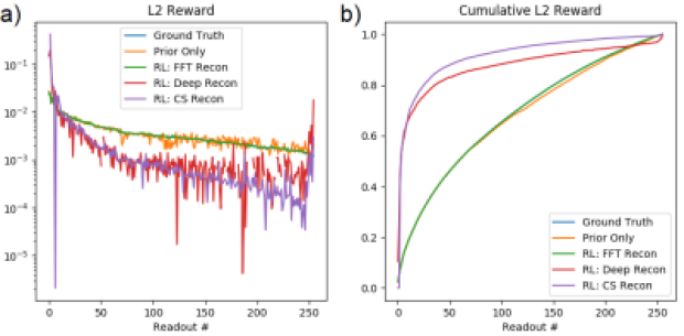
Reinforcement Learning for Online Undersampling Pattern Optimization
David Zeng, Christopher Sandino, Dwight Nishimura, Shreyas Vasanawala, Joseph Cheng
Magnetic resonance imaging (MRI) is an important but relatively slow imaging modality. MRI scan time can be reduced by undersampling the data and reconstructing the image using techniques such as compressed sensing or deep learning. However, the optimal undersampling pattern with respect to image quality and image reconstruction technique remains an open question. To approach this problem, our goal is to use reinforcement learning to train an agent to learn an optimal sampling policy. The image reconstruction technique is the environment and the reward is based upon an image metric.