How Stanford research is making MRI scans safer for children
An interview with radiologist Shreyas Vasanawala

The facade of an MRI machine at Packard Children’s Hospital was made to resemble a sandcastle.
Courtesy of Lucile Packard Children's Hospital
When it comes to medical imaging, pediatric radiologist and biomedical engineer Shreyas Vasanawala knows that kids aren’t the same as adults.
Vasanawala, MD, PhD, professor of radiology at the Stanford School of Medicine, has spent the last 10 years studying how to improve magnetic resonance imaging scans for his smallest, wiggliest patients. Now, he’s putting his MRI innovations to work in the Cynthia Fry Gunn and John A. Gunn Imaging Center at the new Lucile Packard Children’s Hospital Stanford, which opened in December.
Vasanawala talked with Stanford Medicine News about the needs that spurred his inventions and how the new hospital’s state-of-the-art technology will improve his team’s ability to care for children who need medical scans.
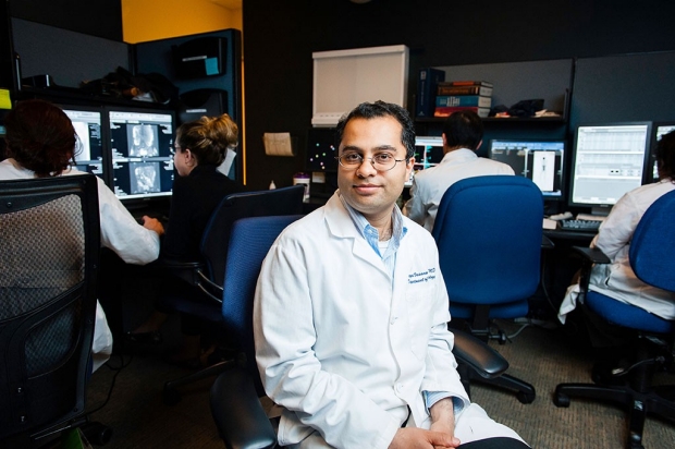
Shreyas Vasanawala tailors MRI equipment to the needs of children.
Toni Bird
Q: MRI scans are noninvasive and painless, don’t use radiation, and give clear images of soft tissue such as muscles and tendons, but children who could benefit from MRIs don’t always get them. Why not?
Vasanawala: Magnetic resonance technology is challenging to develop and use. Most of the MRI equipment on the market was designed to meet the needs of adult patients, who receive about 90 percent of MRI exams.
In an MRI scanner, the body is exposed to a very strong magnetic field. The protons in the body’s water molecules align themselves with the magnetic field. Then, as they return to their usual state, they give off radio-frequency signals detected by the scanner, which translates the signals into a picture.
To produce a clear picture, a traditional MRI scan requires that patients hold very still, sometimes for up to an hour. That’s difficult for young children. Children are also smaller, breathe faster and have higher heart rates — all factors that make the imaging challenges harder from a physics perspective. Kids may be given anesthesia to help them hold still, but that carries its own risks.
Q: As part of your research at Stanford, you’ve been designing MRI equipment especially for children. What improvements have you introduced?
Vasanawala: We’ve invented solutions that have allowed us to eliminate the need for anesthesia in many cases.
We’ve been collaborating with engineers from UC-Berkeley to create new designs and production methods for highly flexible and lightweight MRI signal-receiving coils tailored to children’s bodies. Standard coils are larger than what children need, making them unnecessarily heavy and uncomfortable. Larger-than-necessary coils also pick up extra noise or interference, reducing the image quality. Child-size receiver coils increase image clarity and lower scan times.
The smaller coils greatly enhance the performance of a novel hybrid imaging technology called PET-MR, which we are now offering to patients in our new imaging center at Packard Children’s Hospital. The coils are being developed commercially as well.
Q: You’ve also improved the computing software that processes MRI data. How?
Vasanawala: We’ve created new image-reconstruction algorithms that work better for kids. We deployed motion-
correction strategies that produce sharp images even when a child is moving slightly — this helps address the challenge of kids’ faster heart rates and breathing rates. Simultaneously, to reduce scan times, we implemented novel, high-dimensional imaging and compressed sensing coupled with artificial intelligence. These techniques allow the computer to reconstruct a full MR image from much less raw data. Scans that once took an hour are now complete in five to 10 minutes. This has had a particularly large impact for our cardiac, oncologic and musculoskeletal exams.
Q: What most excites you about the new imaging center at Packard Children’s?
Vasanawala: For the first time, we have an MRI scanner located inside our neurosurgery operating suite. It allows our neurosurgeons to confirm the success of a surgical procedure, such as a tumor resection, before surgery is complete. This saves time by eliminating a separate post-surgical MRI and the risk of needing an immediate repeat surgery. Patients will be spared a second round of anesthesia, hospital stays will be shortened, and families will know if the surgical aims have been achieved as soon as their children are out of the operating room.
By the end of 2019, we will have a next-generation MRI scanner with much stronger magnetic field gradients that can be altered at high speed. This enables faster imaging and better image contrasts. Also, this new MRI scanner will come with a noninvasive technology used to kill certain types of tumors with sound waves. Known as MR-guided high-intensity focused ultrasound, it lets us pinpoint abnormal areas in the body, such as some types of tumors, and heat them to destroy abnormality without cutting into surrounding healthy tissue.