GABAa Receptor Physiology in Autism Spectrum Disorders Including Fragile-X Syndrome
The type A gamma aminobutyric acid receptor (GABAA) is a pentameric transmembrane receptor and a ligand-gated ion channel, which generally situates post-synaptically and mediates inhibitory neurotransmission. Converging evidence suggests that GABAergic dysfunction occurs in both Autism Spectrum Disorder (ASD) and Fragile X Syndrome (FXS). Animal models of ASD as well as postmortem brains of individuals with ASD have shown aberrant GABA physiology. For example, reduced GABAA receptor density has been observed in the hippocampus, cingulate cortex, and fusiform gyrus of the postmortem brains of young adults with ASD. Using an alpha5-selective GABAA receptor radioligand, a pilot study showed that individuals with ASD had lower receptor densities in almost all areas of the brain compared to controls. Lower levels of the GABAA alpha5 subtype were also found in the amygdala and nucleus accumbens bilaterally. A more comprehensive understanding of the GABAergic system in living humans can be achieved by mapping GABAA receptors with PET, GABA with MRS, and structural neuroanatomy with sMRI, co-registering these data, and determining the relationships between these. Although preliminary data on the distribution of GABAA receptor and GABA in ASD are available, no studies have attempted to measure both GABAA receptor and GABA in humans. Further, there have been no attempts to link GABA physiology with neuroanatomy and structural connectivity in ASD and FXS. 11C-flumazenil-PET has been used to study GABAA receptor distribution in other genetic syndromes with autistic features such as Prader-Willi syndrome, Angelman syndrome, and succinic semialdehyde hydrogenase deficiency. Since there is overwhelming evidence supporting the importance of the GABAergic system in ASD and FXS, our goal is to perform clinical investigations of this system using 18F-flumazenil-PET in human ASD and FXS.
More on Fragile X Syndrome
Fragile X syndrome (FXS) is the most common genetic cause of autism spectrum disorder and intellectual disability. Individuals with full mutation lack the FMR1 gene, which codes for the fragile X mental retardation protein (FMRP). FMRP binds to the mRNA of key synaptic proteins, including the delta subunit of the type A gamma aminobutyric acid (GABAA) receptor. The importance of GABAergic dysfunction in FXS has been suggested by studies of Fmr1 knock-out (KO) mice. However, these studies focused on the forebrain while the rest of the brain was largely unexplored. In human studies of FXS, multiple brain networks involving both cortical and sub-cortical areas appear to develop and function in an aberrant fashion. Therefore, our understanding of the developmental dynamics of the GABAergic system in FXS is far from complete. Multimodal neuroimaging can be used as a powerful tool to further understand the role of the GABAergic system in FXS for many reasons. First, visualizing biological constructs in their natural environment (living animal model and human) is clearly the preferred method to study biology. Second, neuroimaging is an ideal tool for studying systems in a longitudinal fashion due to its non-invasive nature. Third, establishing interspecies relationships for any biological parameters between animal models and humans is clearly more achievable with non-invasive techniques such as neuroimaging. Thus, our group is exploring more comprehensively the impact of FMRP deficiency on developmental dynamics of GABAA receptor distribution, GABA levels, structural neuroanatomy and connectional anatomy using multi-modal neuroimaging (i.e., 18F-Flumazenil PET-MR) and pharmacological probing (by oxytocin) in novel animal models of FXS and humans with FXS.
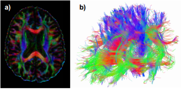
a) Axial image of the brain of a healthy volunteer at 2.5mm isotropic resolution. The colors represent the directions of the underlying fiber pathways (red: left-right, green: anterior-posterior, blue: superior-inferior). B) Fiber tracking based on the diffusion tensor within the same volunteer.
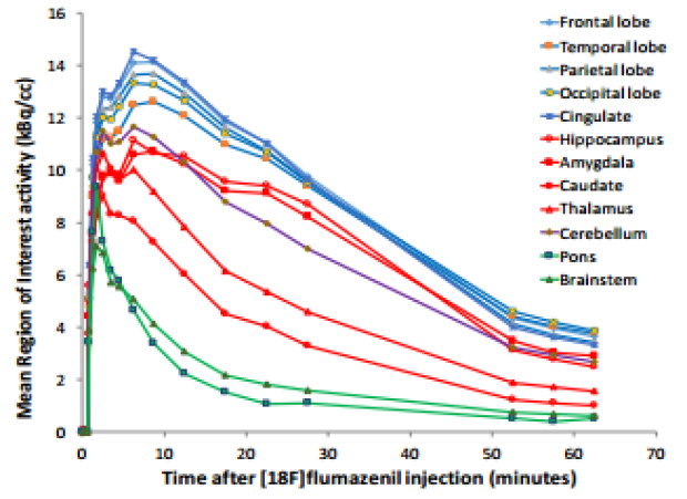
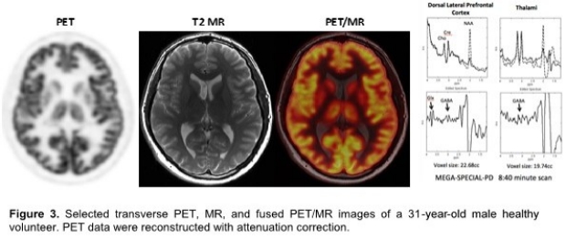
Time-activity curves for regional brain radioactivity after intravenous injection of 5 mCi of [18F]flumazenil in a 31-year-old male healthy volunteer.