MRI of Peripheral Nerves
This work enables high-resolution depiction of nerve fascicular structure - evaluation of such structure can be useful in diagnosing subtle nerve damage.
Ultra-high-resolution MRI with in-plane resolution of about 100um has the potential to improve the visualization of nerve fascicles. However, it is a very challenging task with 3T MRI due to resolution limit imposed by its available signal-to-noise ratio. 7T MRI has the potential for higher resolution than 3T MRI due to the signal boost with higher proton polarization. Our comparison study demonstrated much sharper delineation of nerve fascicular structures can be obtained at 7T MRI than at 3T MRI.
Yoon D, Biswal S, Rutt B, Lutz A, Hargreaves B. Feasibility of 7T MRI for imaging fascicular structures of peripheral nerves. Muscle Nerve. 2018 Mar;57(3):494-8.
This work was featured on the cover of Muscle & Nerve, March 2018.
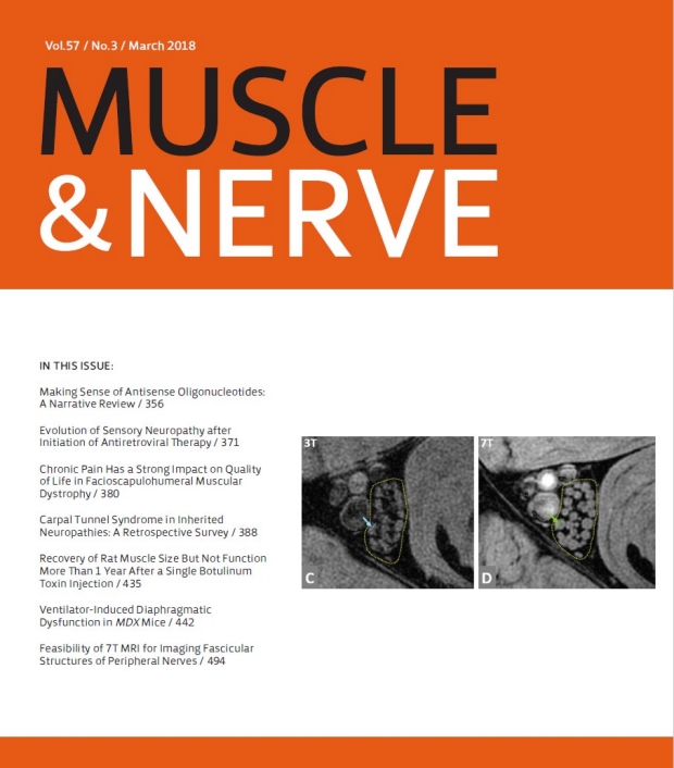
Muscle & Nerve, Journal Cover. March 2018.
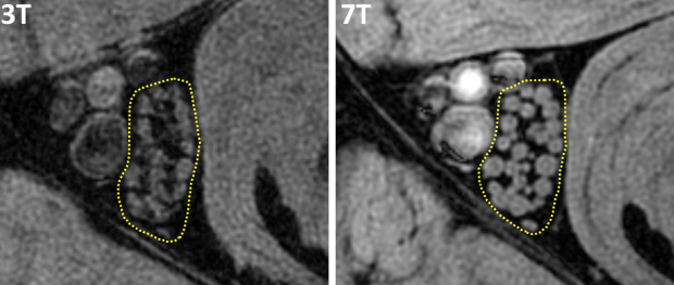
The fascicles of the tibial nerve at high resolution (0.13mm) images acquired at 3T and 7T. Tibial nerve fascicles are enclosed in a dotted line in (C) and (D), and they are more sharply presented at 7T than 3T. Also, the perineurium (the bright rim of each fascicle) is more easily identified at 7T than 3T.