Imaging and T2 Relaxometry of Short-T2 Connective Tissues in the Knee using UTEDESS
Osteoarthritis is a whole-joint disease, but it is currently challenging to image some of the tissues in the knee. This is because tissues such as the menisci, tendons, and ligaments have short-T2 relaxation times which means that they lose energy rapidly following a radiofrequency excitation. More recently, there has been compelling evidence suggesting that measuring the T2 of tissues may be beneficial in assessing their biochemical integrity. Thus, in this study, we propose a new method, termed Ultrashort Echo-Time Double-Echo Steady-State (UTEDESS) which utilizes a 3D-radial k-space acquisition scheme instead of traditional Cartesian sampling. UTEDESS allows imaging and quantification of the T2 of the meniscus, tendons, and ligaments while also providing a retrospective water/far separation as well as images with high-isotropic resolution. This new method is promising for quantitative MRI of the whole knee and may be useful in detecting new biomarkers of osteoarthritis activity.
Chaudhari AS, Sveinsson B, Moran CJ, McWalter EJ, Johnson EM, Zhang T, Gold GE, Hargreaves BA. Imaging and T2 Relaxometry of Short-T2 Connective Tissues in the Knee using Ultrashort Echo-Time Double-Echo Steady-State (UTEDESS). Magn Reson Med. 2017 Dec;78(6):2136-48.
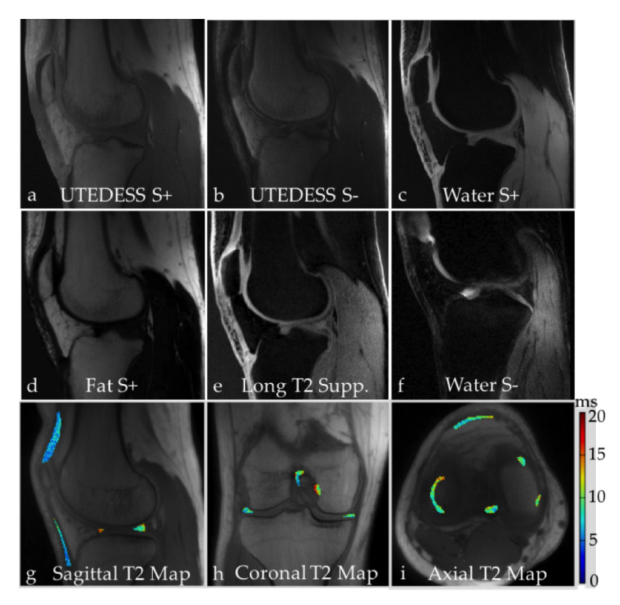
Panels a-f indicate the different contrasts that can be obtained with UTEDESS while panels g-i show how T2 mapping can be performed for short-T2 tissues in arbitrary scan planes.
Bragi Sveinsson and Emily McWalter are alumni of the BMR group