Automatic Renal Segmentation for MR Urography Using 3D-GrabCut and Random Forests
A new, automated workflow uses dynamic MR images of the kidney to segment portions of the kidney and quantitatively characterize regional kidney function.
Chronic kidney disease (CKD), a disease causing gradual loss of kidney function, affects 26 million patients in the US alone. MRI offers the ability to measure regional kidney function, as opposed to “global” kidney function. This, for example, can highlight differences between function of the left and right kidneys that may be important in treatment. Using a combination of automated thresholding and machine-learning techniques, we are able to segment different portions of the kidney (medulla, cortex and collecting system) so that we can characterize the filtration or kidney function.
Yoruk U, Hargreaves BA, Vasanawala SS. Automatic renal segmentation for MR urography using 3D-GrabCut and random forests. Magn Reson Med. 2018 Mar;79(3):1696-707.
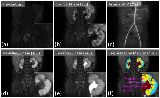
Renal pre-contrast (a) and post-contrast (b, d, e) phases captured by 7-s temporal resolution VDRAD acquisition. (c) The arterial phase can be observed in the maximum intensity projection (MIP) image. Manually drawn segmentation map (f) is used as the ground truth labeling of the renal voxels.
Umit Yoruk is an alumnus of the BMR group