Research
Rejuvenation of aged muscle stem cells and tissues by inhibitng the Gerozyme 15-PGDH
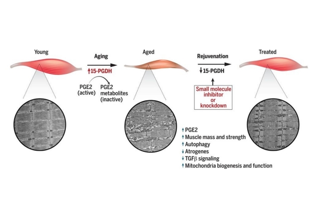
We have discovered that as people age, their muscles accumulate increasing amounts of a protein called 15-PGDH that breaks down a natural inflammatory molecule, Prostaglandin E2 (PGE2), critical to muscle repair after injury. Our data show that reducing the amount of 15-PGDH or using a drug to block its activity in old mice increases their overall health. Treated old mice are able to run longer distances on a treadmill, their muscles are larger, and they are stronger. Blocking 15-PGDH also stimulates muscle stem cells, rejuvenating the muscle’s ability to heal after injury. Conversely, increasing 15-PGDH levels in young mice shrinks and weakens their muscles, mimicking the effect of years of aging in 1 month. These data establish 15-PGDH as a gerozyme, or master regulator of skeletal muscle aging. Our goal now is to understand how changing 15-PGDH abundance causes these effects and if other tissues are similarly impacted. PGE2 helps regulate cell growth and energy production by rejuvenating mitochondria, the cell’s energy source.
Regulation of muscle stem cells in regeneration
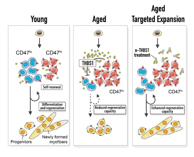
Aging is characterized by a decline in tissue function and regenerative capacity. Sarcopenia, also known as age-dependent loss of skeletal muscle mass and strength, is a major pub-lic-health problem that affects 15% of the elderly, leading to loss of mobility and diminished quality of life. Age-related muscle loss is paralleled by a loss in the function of muscle stem cells (MuSCs), key players in muscle homeostasis and regeneration. However, the mechanisms responsible for age-associated MuSC dysfunction remain elusive. Two major barriers to gaining mechanistic insights into MuSC aging are the heterogeneity of the aged MuSC population, which renders standard bulk analysis ineffective, and the lack of tools to resolve this heterogeneity, underscoring the need for single-cell studies. We previously demonstrated that aged MuSCs are a heterogeneous population comprised of functional and dysfunctional subsets. This key observation suggests a therapeutic strategy to regenerate muscle - boosting the activity of resilient functional MuSCs. We are exploring this possibility using a specific cell surface marker and a series of innovative single-cell technologies required to resolve MuSC subsets.
Reinnervating Muscle
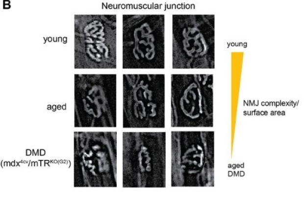
Skeletal muscle can lose innervation with aging, trauma, or neuromuscular disease, leading to weakness and paralysis. Mobility and strength rely on peripheral motor neurons that relay signals from the central nervous system to skeletal muscles. Motor neurons originate in the spinal cord and extend to the skeletal muscles where they branch to innervate multiple myofibers. Each multinucleated myofiber is contacted by a single motor axon at a synapse known as the neuromuscular junction (NMJ). We're investigating how the gerozyme 15-PGDH can be manipulated to promote reinnervation of neuromuscular synapses and recovery of strength after acute or chronic denervation due to injury or aging.
Spinal Muscular Atrophy
Gene therapy has shown remarkable promise for the successful treatment of the root cause of spinal muscular atrophy (SMA). However, combined therapeutic approaches that treat all aspects of SMA, including regenerating muscle, are still urgently needed. We believe that our advances in aging-related muscle wasting and muscular dystrophy can be translated to SMA. We are testing if our novel muscle strengthening therapy, 15-PGDH inhibition, will synergize with an available therapy that restores SMN expression (the root cause of SMA) to increase neuromuscular junctions and improve muscle function and in a severe SMA mouse model. We hypothesize that increasing muscle strength after partial restoration of SMN will provide significant long-term benefits that will improve SMA patient quality of life.
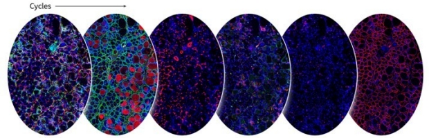
Mapping the muscle stem cell niche
To understand the spatial relationships between the different cell types that make up the muscle, we have optimized multiplexed tissue imaging (CODEX). CODEX allows the identification of cellular neighborhoods. It is ideally suited to study signaling by secreted factors in the local microenvironment because it allows the identification of the cells upon which these factors act. Further, alterations in these relationships can be studied in response to perturbations such as injury, an antibody, or pharmacological intervention. We can visualize lo-calization of up to 60 proteins on single sections of skeletal muscle using antibodies labeled with unique DNA barcodes that are imaged three at time in an iterative process.
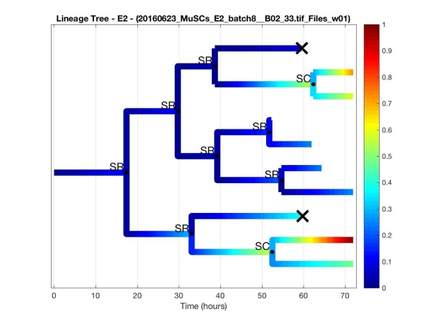
Lineage tracing of stem cell fate decisions
MuSCs undergo asymmetric division. The arsenal of fates a stem cell can access is used by tissues to maintain homeostasis and regenerate thanks to a tightly controlled network of biochemical and biophysical cues. We are developing a dynamic fluorescence time-lapse experiment to evaluate the effects of different conditions on MuSCs fate determination kinetics. Relying on quantified binary signal from the transcription factor Pax7, typical of MuSCs, and myogenin, characteristic of committed myogenic progenitors, we have monitored and classifed division in the three main types and and are comparing different cytokines’ effects on the fate determination scenarios of MuSCs.