Cell Sciences Imaging Facility (CSIF)
About CSIF
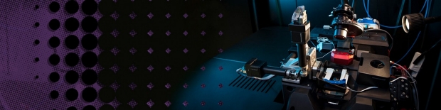
The Cell Sciences Imaging Facility provides high-resolution, state-of-the-art technologies for imaging and analyzing the molecular and structural organization of cells and tissues, as well as bioengineered materials. The facility offers sophisticated and demanding microscopy techniques to Stanford University and industry researchers.
The CSIF is organized into three interdependent imaging labs: the Fluorescence Microscopy Core (FMC), which houses multi-photon, confocal, super-resolution, fluorescence lifetime and deconvolution microscopes, as well as image analysis software; the Electron Microscopy Core (EMC), which houses high-resolution scanning and transmission electron microscopes; and the Spatial Multiplexing Core (SMC), which provides highly multiplexed marker imaging (CODEX) and array tomography services. The CSIF is a participating member of the Stanford Cancer Institute, a National Cancer Institute-designated Comprehensive Cancer Center, supporting cancer research.
In a collaborative effort with Stanford’s School of Engineering (SoE), the CSIF operates a satellite light microscopy facility in the Shriram Center at the SoE. This facility offers biological imaging instrumentation and expertise to the departments of Bioengineering and Chemical Engineering.
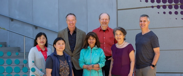
CSIF staff: (front, l-r) Yuanyuan Li, Kitty Lee, Ibanri Phanwar-Wood, Ruth Yamawaki, John Perrino; (back row, l-r) Jon Muholland, David Lenzi

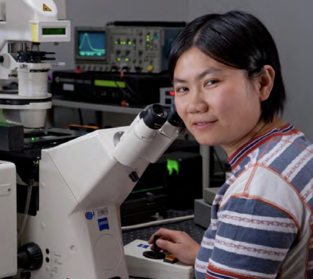
Ibanri Phanwar-Wood with a Leica Ultracut UC7.

John Perrino with a Gatan OneView TEM microscope and OneView 16bit computer.
Services
Light Microscopy
- Confocal microscopy (scanning and spinning-disk)
- Two-photon microscopy
- Wide-field fluorescence microscopy
- Digital deconvolution
- Transmitted-light imaging (phase, DIC, histology)
- High-content screening (confocal and wide-field)
- Super-resolution imaging (SIM and AiryScan)
- Cell surface imaging with <100 nM z-resolution (TIRF)
- Specialized microscopy (FRAP, FRET, FLIP, etc.)
- Fluorescence lifetime imaging microscopy (FLIM)
- Lattice light-sheet microscopy (LLSM)
- Light-sheet microscopy (LSM)
Highly Multiplexed Imaging
- Array tomography
- Akoya CODEX system (50+ biomarkers)
Atomic Force Microscopy
- Bruker BioScope Resolve
Electron Microscopy
- Chemical and cryo-fixation/freeze substitution processing
- High-pressure freezing (w/ opto and electric stimulation)
- Immuno-EM
- Correlative LM to EM: CLEM
- Negative staining
- Ultramicrotomy, cryo and plastic
- Electron tomography (plastic sections)
- High-resolution FE-SEM
- Immuno-SEM
- Array tomography SEM, CLEM
Bioimage Analysis
- Provide software for image analysis, processing, and deconvolution (Imaris, Volocity, SVI Huygens, SoftWoRx, Microvolution)
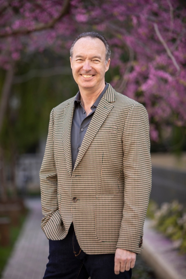
Jon Mulholland
Director, CSIF
SoE Shriram Center and Beckman Center, B050A
jwm@stanford.edu


Yuanyuan Li

Ruth Yamawaki

David Lenzi
Kitty Lee