
Investing in the Beckman Service Centers
By Sarah Williams | The Beckman Center News / Spring 2022
The Beckman Service Centers have a goal of providing our researchers with state-of-the-art scientific technologies. But “state-of-the-art” is a quickly moving target in the biomedical sciences. As quickly as one sequencing technology or imaging device debuts, newer methods and equipment roll out.
To ensure that service center offerings don’t become out of date, the Beckman Center continuously invests in these shared resources. The past year, despite lingering challenges due to COVID-19, was no exception. Here are some of the newest technologies available at our service centers.
Adding New High-Parameter FACS Equipment
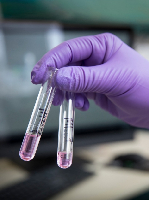
The mainstays of the Beckman Fluorescence Activated Cell Sorting (FACS) Facility include cell sorters, analyzers, and a mass cytometer. For more than 25 years, these pieces of equipment have helped researchers characterize the cellular and molecular details of their biological samples. But with an increased demand for high-throughput, single-cell experiments in recent years, the machines have been more in demand than ever before.
“One of the biggest complaints we’ve had was that it was just too hard to get time on our equipment,” says Lisa Nichols, Ph.D., director of the FACS Facility.
To solve those long waits, Dr. Nichols recently added two new analyzers, a BD Symphony A5 and an Agilent Penteon with an autosampler, each with 5 lasers and up to 30 detectors. To use the new analyzers, researchers can book time themselves, or arrange with the FACS Facility to drop off samples that the service center staff will analyze. And in support of this service, the facility has hired three new staff members.
“Despite inflation and the fact that we’ve added these new high-parameter pieces of equipment and hired new staff, our pricing has remained flat,” points out Dr. Nichols. “And we have an abundance of space on our analyzers, so people can book a day or two out.”
To learn more about how you can use the FACS Facility, visit facs.stanford.edu or contact Dr. Nichols at lisanichols@stanford.edu.
Moving Imaging Technologies Forward
Making sure that the Cell Sciences Imaging Facility (CSIF) is up to date is a full-time job, says Jon Mulholland, director of the CSIF.
“It’s key for me to stay on top of all the new imaging technologies coming out, so our researchers can use the most state-of-the-art approaches to advance their research,” says Mulholland.
Among the most recent technologies added to the CSIF are two new light sheet microscopes—a Zeiss Lattice Lightsheet 7 and a Bruker TruLive3D Imager. The scopes each have unique strengths; the Zeiss excels at live cell imaging with a low photon dose, letting researchers image for hours or days without photobleaching their samples. The Bruker is optimized for imaging small live specimens, including zebrafish and C. elegans.
The CSIF also has pending National Institutes of Health funding for a Leica, Inc., Stellaris DIVE microscope, which would replace a now 12-year-old Leica SP5. Mulholland says users should stay tuned for availability later this year on the new microscope, which offers multicolor imaging in deep tissues.
“These all offer upgrades and new capabilities that are really exciting for our users,” says Mulholland.
For more on the newest imaging technologies, visit microscopy.stanford.edu or email Mulholland at jwm@stanford.edu.
Expanding the Biomolecular Interaction and Spatial Analyses Toolbox
Many people associate the Protein and Nucleic Acid (PAN) Facility with gene expression analyses (next-generation sequencing, microarray, and real-time PCR), oligonucleotide and peptide synthesis, and nucleic acid QC. But the facility offers a large—and growing—array of other tools as well, all designed to study biomolecular interactions and cell states within tissue environments using spatial transcriptomics.
Want to gain insight into, select, and optimize a small molecule compound that you’re interested in during the drug discovery process? The PAN Facility has added another Biacore T200 surface plasmon resonance instrument that can help you characterize the kinetics, affinity, and thermodynamics of an interaction.
Interested in single-cell gene analysis but worried that your cells won’t survive the rough sorting process into a standard 96-well plate? The new BD Rhapsody Single-Cell Analysis System at the PAN Facility is much gentler on cells, says director Michael Eckart, Ph.D. Cells flow over the surface of a cartridge with hundreds of thousands of microwells, and a unique real-time quality control system helps you follow the process as it happens.
Dr. Eckart has also been working to expand the spatial transcriptomics capabilities at the PAN Facility. Today, the facility offers the Visium Spatial Gene Expression solution from 10x Genomics to aid in the analysis of transcriptomics across a whole tissue. Other options are also being explored to expand the toolbox.
“We’re currently evaluating other technologies in that space to determine what would be the best platform to bring on board,” says Dr. Eckart. “We’re interested in hearing from anyone who has an interest in that area and has a technology they believe would be a good option for us to consider.”
That request for feedback is not just limited to spatial transcriptomics, he adds. “I always encourage people to come talk to us about any new technologies they’re considering or that they see coming up on the horizon,” says Dr. Eckart. “We rely on researchers to tell us what is going to help them in achieving their research goals.”
You can reach Dr. Eckart at meckart@stanford.edu or find out more about the PAN Facility at pan.stanford.edu.
Increasing Computing Power
With most new biomedical technologies also comes an increased need for software, data storage, and computing power to handle data and analysis. The Computational Services and Bioinformatics Facility (CSBF) this year installed new Windows servers to host a variety of software for proteomics research as well as the VisioPharm software used by CSIF. They are also providing new software support to the FACS Facility with the addition of FCS Express, which is used to analyze data from cell sorting machines.
Lee Kozar, director of CSBF, encourages researchers of all levels to contact him when they have new software needs, to determine if the facility can help arrange licensing agreements. He can be reached at kozar@stanford.edu; more information about CSBF is at cmgm-new.stanford.edu/csbf/.
| Instrument | Service Center | Manufacturer |
| OneView Scintillator-Coupled sCMOS Transmission Microcsope (TEM) | CSIF |
Gatan, Inc. |
| CODEX microfluidics highly multiplexed imaging platform | CSIF |
AKOYA Biosciences, Inc. |
| Ultracut UC7 ultramicrotome | CSIF |
Leica Microsystems, Inc. |
Luxendo TruLive 3D Imager |
CSIF |
Bruker Nano, Inc. |
| LSM 900 Confocal | CSIF |
Carl Zeiss Microscopy, LLC |
| FACSymphony 5-laser, 30 parameter analyzer | FACS |
BD Biosciences |
| 5-Laser Agilent Penteon Analyzer | FACS |
Agilent Technologies |
5-Laser, 29-color FACSymphony |
FACS |
BD Biosciences |
| 50-parameter FACSymphony S6 Spectral Sorter | FACS |
BD Biosciences |
| Server to support multiplex imaging technology | CSBF |
Colfax International |
| Biacore T200 Instrument | PAN |
Global Life Sciences Solutions USA, LLC |
For more information (media inquiries only), contact:
Naomi Love
(650) 723-8423
naomi.love@stanford.edu
More Beckman Center News | Spring 2022
Sign up to receive our quarterly newsletter
