Two Generous Grants Boost CODEX Imaging Capabilities
By Sarah Williams | The Beckman Center News / Winter 2022
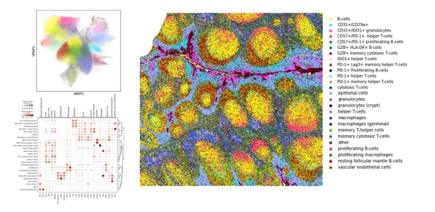
Spatial phenotyping with Akoya Bioscience's CODEX (now PhenoCycler) platform.
Two new grants to Beckman Center service centers expand researchers’ access to highly multiplexed tissue imaging.
So you have a tissue sample, and dozens of proteins that you want to pinpoint the location of; what do you do? Classic fluorescence microscopy lets you visualize only a few colored tags at a time, but CODEX—short for CO-Detection by indEXing—will allow you to simultaneously look at many more tags.
Thanks to two new grants, the Beckman Center service centers recently expanded their ability to support CODEX imaging—with new automated sample preparation, imaging equipment, and artificial intelligence software that analyzes results.
“There’s been a lot of interest in this technology, and we were really backlogged with just one set of equipment,” says Anum Khan, the senior staff scientist who runs the platform at the Cell Sciences Imaging Facility (CSIF). “The new grants enabled us to expand very quickly.”
The CODEX multiplex imaging platform relies on antibodies conjugated to DNA barcodes, rather than directly to fluorescent markers. Instead, fluorescent tags are targeted to the barcodes and can be successively bound and washed off, allowing multiple rounds of imaging—and the visualization of up to 40 or 50 different markers—in one single tissue.
The technology is a boon to anyone who wants to study the spatial organization of molecules, says CSIF director Jon Mulholland. Immunologists, cancer researchers, and neuroscientists are among the researchers who use CODEX to get new insights into the complexity of tissues and disease.
A c-ShARP Grant
The first grant for the imaging approach was awarded last summer. CSIF was awarded $268,000 through the Community of Shared Advanced Research Platforms (c-ShARP) funding program, a new university-wide campus initiative operated by the office of the University Dean of Research. C-ShARP supports shared facilities across Stanford, with the aim of encouraging collaborative, multidisciplinary research with state-of-the-art instrumentation.
At CSIF, the funds were used for a second PhenoCycler system—the instrument previously known as CODEX and required for the multiplex imaging technology—as well as a new Keyence BZ-X810 microscope, Visiopharm image analysis software, and Parhelia Biosciences and Opentrons automated tissue staining devices.
“This really streamlines our processes and makes things easier for our users,” says Mulholland.
An Anonymous Grant
The second grant, which was made anonymously, awarded $17,000 to Beckman’s Computational Services and Bioinformatics Facility (CSBF), to support the server needed for the expanded imaging platform.
“This server is ten years newer than what we had before; it has more memory, more disk space, and a faster processor,” says CSBF director Lee Kozar. “On our old system, we couldn’t run all these new software packages.”
The new imaging instrumentation and software have been up and running since the fall, and both CSIF and CSBF welcome new users.
Of course, demand for multiplex imaging remains high. Khan and Mulholland are already eyeing the next generation of PhenoCycler, which would speed up the workflow even more.
For more information on multiplex imaging, and advice on how best to image your samples, contact Jon Mulholland (jwm@stanford.edu) or Anum Khan (anumkhan@stanford.edu) at the Cell Sciences Imaging Facility.
For more information (media inquiries only), contact:
Naomi Love
(650) 723-8423
naomi.love@stanford.edu
More Beckman Center News | Winter 2022
Sign up to receive our quarterly newsletter