Research Tools

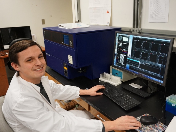
Mass Cytometry
Mass cytometry, or CyTOF (Fluidigm), is a variation of flow cytometry in which antibodies are labeled with heavy metal ion tags rather than fluorochromes. Readout is by time-of-flight mass spectrometry. This allows for the combination of many more antibody specificities in a single samples, without significant spillover between channels. Mass cytometry technology allows us to enable deep profiling of translational and clinical research samples across a range of cell surface and intracellular markers. This allows us to identify the biomarkers and discover underlying immune mechanisms for allergy and asthma research.
Flow Cytometry
Flow cytometry is a widely used method for analyzing the expression of cell surface and intracellular molecules, characterizing and defining different cell types in a heterogeneous cell population, assessing the purity of isolated subpopulations and immune cell functions. It allows simultaneous multi-parameter analysis of single cells. Our FACScan Analyzer has been upgraded to 4-laser, 10-color configuration. Its current laser/detector configuration provides 4-Violet, 2-Red, and 4-Blue laser excited detection channels. This technique allows us to identify the biomarkers and discover underlying immune mechanisms for allergy and asthma research.
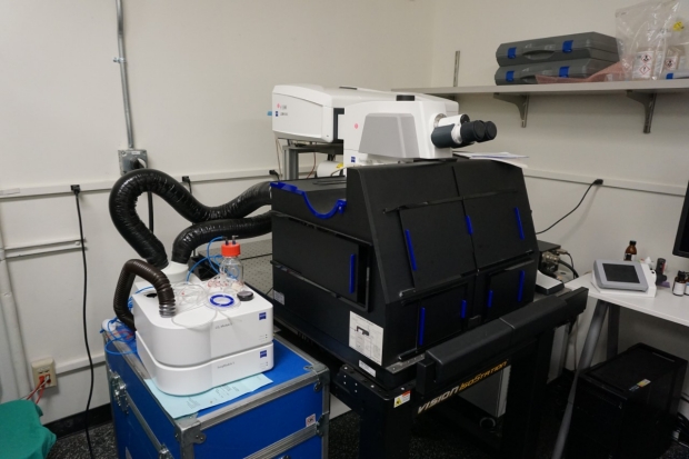
Confocal Microscopy
Zeiss 880 Examiner super resolution confocal microscope allows us to image an extremely broad spectrum of live fluorescent proteins, in deep tissue, with super resolution. The Center has been using this microscope to study how stem and progenitor cells that are normally responsible for repair and regeneration of the airways in healthy individuals become mis-programmed in asthma to lead to airway hypersensitivity. Using the unprecedented imaging this microscope makes possible, we have discovered previously unappreciated changes to blood vessels in asthmatic lungs and we are currently investigating the cells and molecules responsible and how those vessel changes impact pulmonary physiology. Zeiss designed and built this imaging system specifically for our needs, and it is an absolutely indispensable tool for our studies that has led to very exciting new discoveries.
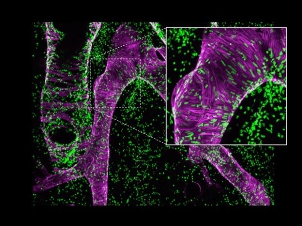
An airway (magenta, left side) and artery (magenta, right side) where the nuclei of proliferating cells have been marked in green; the inset is a higher magnification view of the artery wall.

A pulmonary artery in which individual cells in the vessel wall have been labeled with blue, orange or red fluorescent proteins. Magenta is smooth muscle cells surrounding the airways (behind the artery) and the artery.

A whole lobe of a transgenic mouse lung, with red, orange, and yellow marking the smooth muscle and green marking the nerves.
HiSeq 4000 Sequencing Systems
The HiSeq 4000 Systems leverage innovative patterned flow cell technology to provide rapid, high-performance sequencing. Perform production-scale, high-throughput exome or transcriptome sequencing projects quickly and economically. We use the system as part of biomarker discovery in food oral immunotherapy clinical trials.
Photos from our Research Projects
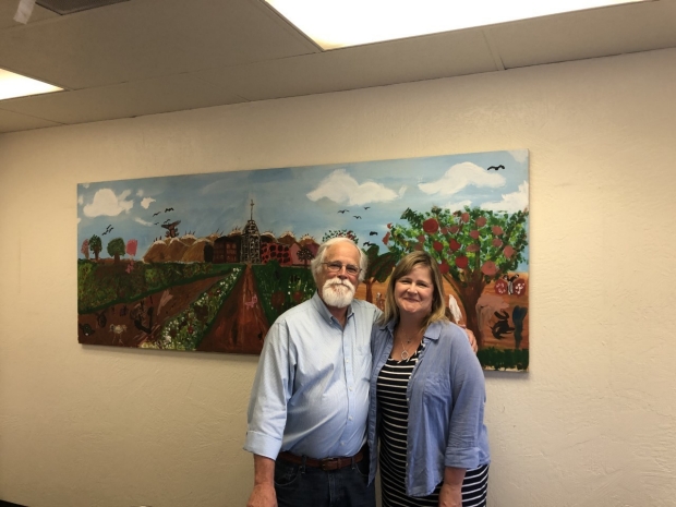
Our work with the Fresno community

Our work with the Stanford Proteomics and Metabolomics Facility