Research
Our lab takes an interdisciplinary approach to develop novel therapeutics and surgical techniques for the treatment of heart failure and structural heart disease. With experience in molecular and cellular biology, tissue engineering, biomaterials engineering, mechanical engineering, radiology, and surgery, we test potential therapies, medical devices, and surgical repair techniques in vitro or ex vivo, then in vivo in small and large animal models, and ultimately in humans.


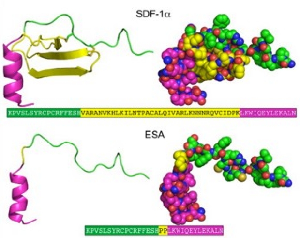
We have studied the potent chemokine stromal cell-derived factor 1α (SDF) and its role in activating angiogenesis and myocardial repair mechanisms after myocardial infarction. Via computational protein chemistry, we then designed and synthesized a supra-efficient engineered SDF analog (ESA). When delivered after myocardial infarction, ESA augmented endogenous angiogenesis that translated to borderzone preservation and cardiac functional improvements in both small and large animal models. We were also able to significantly improve local tissue biomechanical properties and ventricular geometry after myocardial ischemic injury.

Selected Publications
Hiesinger W, Perez-Aguila JM, Atluri P, et al. Computational protein design to reengineer stromal cell-derived factor-1α generates an effective and translatable angiogenic polypeptide analog. Circulation. 2011; 124:S18-26.
MacArthur JW Jr, Cohen JE, McGarvey JR et al. Preclinical evaluation of the engineered stem cell chemokine stromal cell-derived factor 1α analog in a translational ovine myocardial infarction model. Circ Res. 2014; 114:650-659.
Goldstone AB, Burnett CE, Cohen JE, et al. SDF 1-alpha attenuates myocardial injury without altering the direct contribution of circulating cells. J Cardiovasc Transl Res. 2018; 11:274-278.
Wang H, Wisneski A, Paulsen MJ, et al. Bioengineered analog of stromal cell-derived factor 1α preserves the biaxial mechanical properties of native myocardial after infarction. J Mech Behav Biomed Mater. 2019; 96:165-171.
Our laboratory has investigated the potential of various molecular therapies to induce myocardial regeneration after ischemic injury, including cell cycle regulators and neuregulin. We have also studied the reparative paracrine effects of mesenchymal stem cells, which led to a phase 1 multicenter, prospective, randomized, placebo-controlled, double-blinded human clinical trial that demonstrated efficacy, and phase 2 trial that was recently completed.
We have also studied the remarkable ability of neonatal mammals to naturally regenerate the heart after mechanical injury or myocardial infarction. Using microsurgical techniques and transgenic mice to perform cellular lineage-tracing experiments, we demonstrated the importance of neovasculogenesis and collateral artery formation in the process of natural heart regeneration. In addition to mice, we have also demonstrated that newborn rats are capable of neonatal heart regeneration as well, resulting in negligible scar formation after myocardial infarction and complete preservation of normal cardiac geometry and function.


Selected Publications
Woo YJ, Panlilio CM, Cheng RK, et al. Therapeutic delivery of cyclin A2 induces myocardial regeneration and ehnahces cardiac function in ischemic heart failure. Circulation. 2006; 114:I206-213.
Cohen JE, Purcell BP, MacArthur JW Jr, et al. A bioengineered hydrogel system enables targeted and sustained intramyocardial delivery of neuregulin, activating the cardiomyocyte cell cycle and enhancing ventricular function in a murine model of ischemic cardiomyopathy. Circ Heart Fail. 2014; 7:619-626.
Ascheim DD, Gelijns AC, Goldstein D, et al. Mesenchymal precursor cells as adjunctive therapy in recipients of contemporary left ventricular assist devices. Circulation. 2014; 129:2287-2296.
Ingason AB, Goldstone AB, Paulsen MJ, et al. Angiogenesis precedes cardiomyocyte migration in regenerating mammalian hearts. J Thorac Cardiovasc Surg. 2018; 155:1118-1127.
Das S, Goldstone AB, Wang H, et al. A unique collateral artery development program promotes neonatal heart regeneration. Cell. 2019; 176:1128-1142.
Wang H, Paulsen MJ, Hironaka CE, et al. Natural heart regeneration in a neonatal rat myocardial infarction model. Cells. 2020; 9.
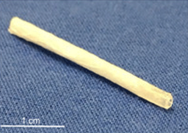
We are investigating the benefit of novel protein cytokine therapeutics delivered to the myocardium in translatable hydrogels. By encapsulating cytokines within injectable hydrogels, we have demonstrated improved therapeutic localization and significantly prolonged cytokine half-lives in vitro and in vivo, ultimately optimizing the temporospatial window in which these cytokines are active. Our most recent hydrogel features two distinct release mechanisms, thereby allowing the delivery of two different cytokines each at a different rate tailored to the properties of the individual cytokine. Moreover, this hydrogel is also able to be delivered to the heart through a long 4 Fr catheter, thus permitting a percutaneous treatment strategy for future clinical translation.
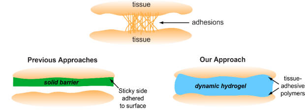
Another application for viscoelastic materials is the prevention of post-operative adhesions. In cardiac surgery, pericardial adhesions are particularly problematic during reoperations, as surgeons must release the adhesions from the surface of the heart before the intended procedure can begin, thereby substantially lengthening operation times and introducing risks of hemorrhage and injury to the heart and lungs during sternal re-entry and cardiac dissection. We have shown that shear-thinning, self-healing hydrogels dramatically reduce the severity of pericardial adhesions in small and large animal models, presenting a promising clinical solution for the prevention of post-operative adhesions.
Selected Publications
MacArthur JW Jr, Purcell BP, Shudo Y, et al. Sustained release of engineered stromal cell-derived factor 1α from injectable hydrogels effectively recruits endothelial progenitor cells and preserves ventricular function after myocardial infarction. Circulation. 2013; 128:S79-86.
Steele AN, Stapleton LM, Farry JM, et al. A biocompatible therapeutic catheter‐deliverable hydrogel for in situ tissue engineering. Adv Healthc Mater. 2019; 8:e1801147.
Stapleton LM, Steele AN, Wang H, et al. Use of a supramolecular polymeric hydrogel as an effective post-operative pericardial adhesion barrier. Nat Biomed Eng. 2019; 3:611–620.
Steele AN, Paulsen MJ, Wang H, et al. Multi-phase catheter-injectable hydrogel enables dual-stage protein engineered cytokine release to mitigate adverse left ventricular remodeling following myocardial infarction in a small animal model and a large animal model. Cytokine. 2020; 127:154974.
We are studying a unique cell culture technology that produces cell sheets with naturally-produced extracellular matrix and intact cell-cell interactions. These sheets have excellent handling characteristics and can be implanted onto the heart, thereby delivering cells while minimizing dispersion, promoting natural cellular interactions, and enhancing cell survival. To better mimic the architecture of a mature blood vessel, we have created bilevel cell sheets composed of endothelial progenitor cell (EPC) and smooth muscle cell (SMC) layers to enhance survival of transplanted cells, to effect angiogenesis after myocardial infarction, and to study the fate of transplanted EPCs.
We have also designed and engineered a bilayer vascular conduit using SMC-fibroblast bi-level cell sheets. This engineered vessel was successfully utilized as a femoral interposition graft in a rodent model. We are currently upscaling the engineered vascular conduit using cell sheets from a more translatable source (bone marrow and peripheral blood rather than aorta) to test in a preclinical large animal model. This will allow us to better realize the capacity of cell sheet technology for vasculogenesis with cellular therapeutics.

Selected Publications
Shudo Y, Cohen JE, MacArthur JW, et al. Spatially oriented, temporally sequential smooth muscle cell-endothelial progenitor cell bi-level cell sheet neovascularizes ischemic myocardium. Circulation. 2013; 128:S59-68.
Shudo Y, Goldstone AB, Cohen JE, et al. Layered smooth muscle cell-endothelial progenitor cell sheets derived from the bone marrow augment postinfarction ventricular function. J Thorac Cardiovasc Surg. 2017; 154:955-963.
Kawamura M, Paulsen MJ, Goldstone AB, et al. Tissue-engineered smooth muscle cell and endothelial progenitor cell bi-level cell sheets prevent progression of cardiac dysfunction, microvascular dysfunction, and interstitial fibrosis in a rodent model of type 1 diabetes-induced cardiomyopathy. Cardiovasc Diabetol. 2017; 16:142.
von Bornstädt D, Wang H, Paulsen MJ, et al. Rapid self-assembly of bioengineered cardiovascular bypass grafts from scaffold-stabilized, tubular bilevel cell sheets. Circulation. 2018; 138:2130-2144.
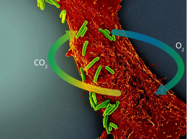
The core problem underlying ischemic injury is insufficient delivery of oxygen, glucose, and other essential molecules to tissues, combined with simultaneous buildup of carbon dioxide that cannot be cleared. To permit aerobic respiration to continue in the setting of tissue ischemia, we have successfully employed a photosynthetic strategy in which cyanobacteria are introduced to the heart after myocardial infarction. These photosynthetic organisms capture carbon dioxide from ischemic cells and water from the extracellular environment and recycle them into oxygen and glucose in the presence of light. In a rat ischemia/reperfusion model, treatment with cyanobacteria yielded a 25-fold increase in tissue oxygen levels and improved cardiac output by nearly 60% compared to untreated, ischemic controls.
Selected Publications
Cohen JE, Goldstone AB, Paulsen MJ, et al. An innovative biologic system for photon-powered myocardium in the ischemic heart. Sci Adv. 2017; 3:e1603078.
Wang, H, Wu MA, Woo YJ. Photosynthetic symbiotic therapy. Aging. 2019;11:843-844.
Advancements in Ex Vivo Simulation and Disease Modeling
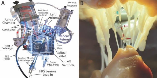
Using our department's high-resolution 3D printing facilities, we designed and manufactured a custom left heart simulator that generates physiologic pressures and flows through the mitral and aortic valves. We are continually working to advance the capabilities of cardiac biomechanics simulation while developing novel measurement technologies. To overcome limitations in previous strain gauge measurements of chordae tendineae, we created custom force-sensing neochordae using Fiber Bragg Grating technology that mimic the natural shape and movement of native chordae. Due to their lightweight and thin-profile design, these sensors allow us to measure chordal forces without disrupting the hemodynamics and structural integrity of the mitral valve.
Furthermore, we have leveraged interdisciplinary engineering approaches and novel device design to successfully model a variety of different pathologies, including Barlow's mitral valve using a cross-species model with a bovine valve implanted within a porcine-sized annulus, and mitral annular dilation using an iris-inspired device.

Selected Publications
Zhu Y, Imbrie-Moore AM, Paulsen MJ, et al. A Novel Aortic Regurgitation Model From Cusp Prolapse With Hemodynamic Validation Using an Ex Vivo Left Heart Simulator. J Cardiovasc Transl Res. 2020 Jun 3. doi: 10.1007/s12265-020-10038-z.
Paulsen MJ, Bae JH, Imbrie-Moore A, et al. Development and ex vivo validation of novel force-sensing neochordae for measuring chordae tendineae tension in the mitral valve apparatus using optical fibers with embedded Bragg gratings. J Biomech Eng. 2019; doi:10.1115/1.4044142.
Imbrie-Moore AM, Paullin CC, Paulsen MJ, et al. A novel 3D-printed preferential posterior mitral annular dilation device delineates regurgitation onset threshold in an ex vivo heart simulator. Med Eng Phys. 2020; 77:10-18.
Imbrie-Moore AM, Paulsen MJ, Zhu Y, et al. A novel cross-species model of Barlow’s disease to biomechanically analyze repair techniques in an ex vivo left heart simulator. J Thorac Cardiovasc Surg. 2020 (in press).
Surgical Repair Optimization
Using our ex vivo heart simulator, we are actively studying how surgical repair techniques influence the biomechanics of the aortic and mitral valves. Armed with this data, we aim to optimize repairs for long-term durability and also develop novel devices and approaches to improve cardiac surgery. We have published an ex vivo analysis comparing two of the conduit choices available for valve-sparing aortic root replacement—the straight graft and the Valsalva graft. Additionally, we have studied a variety of different repair techniques for mitral regurgitation including apical neochord anchoring and the non-resectional posterior ventricular anchoring nechordal (PVAN) repair—a technique that was developed by Dr. Woo.

Selected Publications
Paulsen MJ, Kasinpila P, Imbrie-Moore AM, et al. Modeling conduit choice for valve-sparing aortic root replacement on biomechanics with a 3-dimensional-printed heart simulator. J Thorac Cardiovasc Surg. 2018; 158:392-403.
Imbrie-Moore AM, Paulsen MJ, Thakore AD, et al. Ex vivo biomechanical study of apical versus papillary neochord anchoring for mitral regurgitation. Ann Thorac Surg. 2019; 108:90-97.
Paulsen MJ, Imbrie-Moore AM, Wang H, et al. Mitral chordae tendineae force profile characterization using a posterior ventricular anchoring neochordal repair model for mitral regurgitation in a three-dimensional-printed ex vivo left heart simulator. Eur J Cardiothorac Surg. 2020; 57:535-544.
With access to recently-renovated small animal operating suites, a state-of-the-art hybrid large animal operating theater supporting both endovascular and open surgery, and a 3T MRI, the Woo Lab possesses extensive experience with testing a variety of cardiovascular therapies in preclinical animal models. Working in collaboration with the Stanford Veterinary Services Center, our laboratory has also developed various novel translational animal models to facilitate the testing of potential promising therapies.

Dr. Woo emphasizes the importance of research in advancing the field of cardiovascular surgery. He has served as the local principal investigator for multi-center clinical trials that test the newest cardiac surgical devices, ranging from sutureless valve prostheses to mechanical circulatory support devices (heart pumps). Additionally, therapies conceived in the laboratory are ultimately investigated for safety and efficacy in humans. Dr. Woo has been at the frontier of cell therapy for the surgical treatment for heart failure. He has tested direct endothelial progenitor cell injection during coronary artery bypass surgery and served as the lead principal investigator of the Cardiothoracic Surgical Trials Network's (CTSN) investigation of the safety and efficacy of intramyocardial injection of mesenchymal precursor cells on myocardial function in LVAD recipients. Given initial success, Dr. Woo and the CTSN received additional funding and recently completed the phase II trial of mesenchymal precursor cells for the treatment of heart failure.
Therapies that are effective in small animal models often fail to demonstrate utility in humans. Furthermore, animal models do not exist for a number of idiopathic diseases that affect the heart. Thus, our lab, in collaboration with Euan Ashley’s lab, is building a large biorepository of human cardiovascular tissue (both diseased and healthy). The biorepository exists to help investigators obtain the tissue necessary for gaining mechanistic insight into human cardiovascular diseases as well as test potential therapies. If interested in obtaining human tissue for cardiovascular research, please see the Contact Us page.