Astrid Gillich, PhD
Episode 1: Basic Science Approach with Ross Metzger, PhD & Astrid Gillich, PhD
This new podcast series comes to you from The Vera Moulton Wall Center for Pulmonary Vascular Disease at Stanford, with the goal of addressing specific research efforts, educational programs, and advancements in treatment and patient care.
Synopsis:
In the first in a 3-Part Series on Ways to Attack Pulmonary Vascular Disease, Stanford researchers Astrid Gillich, PhD and Ross Metzger, PhD explore the basic science approach and discuss their discovery of two capillary cell types.
Host:
Welcome to the PH at Stanford Podcast. This new podcast series comes to you from the Vera Moulton Wall Center for Pulmonary Vascular Disease at Stanford, with the goal of addressing specific research efforts, educational programs, and advancements in treatment and patient care.
Today, is the first in a three-part COVID-related series on Ways to Attack Pulmonary Vascular Disease. Stanford researchers, Astrid Gillich, PhD and Ross Metzger, PhD, will be giving us a closer look at our lungs, discussing their discovery of two capillary cell types, as they explore the basic science approach of their research.
Astrid Gillich, PhD:
Hello, everyone. My name is Astrid Gillich. I'm a basic scientist at Stanford University. I am part of the Wall Center for Pulmonary Vascular Disease and today I am very excited to talk to you about our work on the blood vessels of the lung.
Ross Metzger, PhD:
Hi, I'm Ross Metzger. I am also a researcher doing basic research in the Wall Center at Stanford. I began studying the lung more than 20 years ago as a graduate student in the lab of Mark Krasnow who's now the executive director of the Wall Center. And the work that we're going to tell you about is a collaboration between my lab and Mark's.
Astrid Gillich, PhD:
Our lung is really important, because it functions to bring oxygen into our body, which we need for our cells to survive and function properly. The lung has an extremely complicated architecture to accommodate a really large surface, about half the size of a tennis court. With every breath we take, air enters our lung and travels through a series of branched tubes, and there are literally millions of them, to the interior of the organ, where it reaches tiny air sacs called alveoli. Here, oxygen is transferred across an extremely thin membrane into the blood, and is carried by red blood cells to every part of our body. We refer to this process where oxygen moves into the blood and carbon dioxide is eliminated as gas exchange. The alveoli are the sites of gas exchange.
So what is an alveolus?
An alveolus is essentially a tiny pocket with an opening in walls that are extremely thin to allow efficient transfer of oxygen into the blood. The pocket is made up of cells. These are epithelial cells and we know that there are two types, and they have very distinct structures and functions. Each of these pockets is surrounded by a network of tiny blood vessels, the capillaries. The capillaries are tubes, they are composed of endothelial cells, so they are the cells that make up the walls of the vessels and they are filled with blood. The two layers of cells, the endothelial cells of the capillaries, and the epithelial cells of the alveolus are closely aligned to form the air-blood barrier. The structure of the air-blood barrier is altered and gas exchange is compromised in many different lung diseases, including acute diseases, chronic diseases, and including COVID-19.
Ross Metzger, PhD:
Alveoli were discovered in the 17th century by Marcello Malpighi in Bologna in Italy. Malpighi was using the microscopes of his time when he made these remarkable drawings of the alveoli. What he was really fascinated by, and you can really see this in the drawings, is the architecture of the alveoli, this remarkable structure of the lung. He was really the first to appreciate that. He was interested in trying to understand from what he could see what he could learn about the function of the lung. Malpighi was not only the first to discover the alveoli, but he was the first to discover and he also drew the capillaries surrounding these pockets and that work really inaugurated lung biology, basic research into understanding the structural basis of lung function.
Of course, since Malpighi, there have been remarkable advances. We have a great understanding of the physiology of the lung. A lot of this has been made possible by technologies that didn't exist in the 17th century. Even the structure of the air-blood barrier, the cellular basis for gas exchange, has been really carefully worked out. These are discoveries that have saved lives, and these are the discoveries that can now be found in the textbooks.
When we started our work, the alveolar capillaries, on the blood side of the air-blood barrier, had been much less well studied than the epithelial cells on the air side. According to this textbook account of the lung, there's a single capillary cell type in the alveolar capillaries, and that in fact is thought to be true for capillaries or really blood vessels throughout our bodies. In blood vessels people had thought that cells that sit next to each other were the same cell type, and that's different from what people already knew about the air side of the alveoli, where there are these two different cell types. There was the idea that there was a single cell type, and there was also the idea that the capillaries around the alveoli, though they were somewhat different from capillaries elsewhere in the body were not all that different from capillaries elsewhere in the body.
Astrid Gillich, PhD:
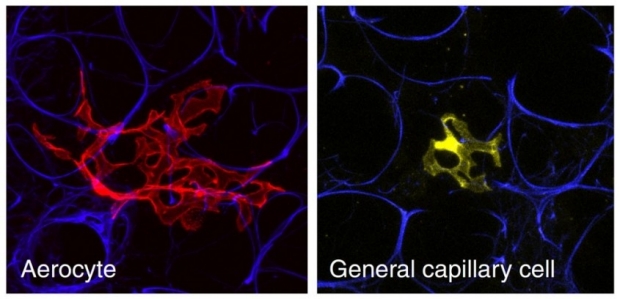
When Ross and I looked really carefully at the blood vessels of the lung, we found that the capillaries are in fact a mosaic composed of two cell types. These cell types are intermingled, but they're really different in their structure and the functions. One of them is really an amazing cell. It is a huge complex cell that has pores, it looks like Swiss cheese, and this cell is so large that it can span multiple alveoli. This cell is specialized in gas exchange, and it is unique to the lungs. We call them aerocytes.
The second cell type is a smaller simpler cell. This cell has another surprising function. These cells function as stem cells in the repair of the capillaries. So they can make more of each of the two capillary cell types.
One of the things that turned out to be really important for us was to look at single cells so that we could really see what they look like since the air-blood barrier is so thin that if you look at all cells you can't really appreciate their morphologies, and their locations, and how they fit together. So that was one of the things. The second key advancement is sequencing technology, so that we can actually look at the diversity of cells and see which of them are molecularly distinct, which of them are different to each other.
Ross Metzger, PhD:
Even this discovery that Astrid mentioned of the two epithelial cell types -- that's relatively recent. It was only in the last 60 or so years, and that discovery was really made possible because of electron microscopy. The lining of these alveoli is so thin that people really didn't think there was a continuous lining. People used to describe the alveoli as an open wound where the tubes just ended, air dumped out into something that had all these blood vessels kind of stuck in it. But with electron microscopy, they could see that there was a continuous lining, and then went on to use other kinds of techniques to discover that there were two cell types. But those two cell types were maybe, in some sense, a little easier to see than what turned out to be the capillary cell types.
So I do think that there is an element where we were able to use a bunch of very new tools without knowing that we were looking for this, to uncover diversity and heterogeneity. Then from there, to do a lot of experiments to get at the function. These powerful new tools would really make it possible to learn a lot about function, and they are going to be really critical tools for starting to understand more about disease.
In general, our approach to disease is to start with cells. We think of diseases as cells doing things that they shouldn't be doing. Our goal is to understand how cells change in disease, and how we can modify the behaviors of the cells to treat the disease, to restore function, or even to regenerate lung tissue. So for that, it's really important, as Astrid said, to understand the full diversity of all the cell types in the lung, and then we can ask how they change in disease.
This is really an approach shared by many researchers in the Wall Center, applying it not just to diseases that affect the gas exchange surface or the capillaries in particular, but really all lung diseases. Just in the last year, Mark Krasnow's lab in collaboration with the Chan Zuckerberg Biohub published a complete cell atlas describing all the cell types in the human lung with molecular information at a really unprecedented level for all of these cell types.
Another Wall Center researcher, Maya Kumar, identified the cell of origin for the occlusive vascular lesions seen in pulmonary arterial hypertension. Thinking about what cell types are there, what they do, and how we can modulate specific cell types is something that these new approaches, and the support of the Wall Center has really made possible.
Astrid Gillich, PhD:
Our findings are really timely now since the alveolus is the site of lung injury induced by viruses, including SARS-CoV-2. The new coronavirus infects the cells of the alveolus and causes damage to the epithelium and the underlying blood vessels. That leads to, essentially, flooding of the alveoli with fluid so the airspaces get filled with fluid. This impairs gas exchange, and it can lead to severe complications and even death from respiratory failure. So what roles the capillary cell types play in COVID-19, and how they are altered is something we really want to explore.
For example, you could imagine that changes in how the capillary cell types interact with other cell types like the immune cells could trigger or initiate inflammation. Or that initiation of coagulation by the capillary cell types could lead to the blood clots that we see in COVID-19 patients. How the capillary stem cells respond to damage, and how we could activate them to trigger the repair of the capillaries is a really important question. It's really that now that we know that there are two cell types that are intermingled in the alveolus, there are suddenly all these questions that we now can explore, and it's really relevant when thinking about disease.
Ross Metzger, PhD:
One of the really exciting things about what we found at this really critical time is that these functions that Astrid just talked about, interactions with other cells, coagulation, these are things that we can see, from what we've learned about the two cell types, are functions that are split up among the cell types.
So coagulation -- each of these cell types plays different roles, and that one cell type has a major role in interacting with the immune cells that's not a function of the other cell type. So some of these functions that we really know are impaired in COVID and really responsible for contributing to the disease, are functions of these specific cell types. So now it's possible to learn much more about what's happening and hopefully do something about it.
Astrid Gillich, PhD:
There's also a lot of interactions between the two cell types, and signaling and communication between them. So you might imagine if you lose one of them that might also impact the other one, and that's also a very interesting question to explore: how do cell types communicate with each other, and how changes in the communication might lead to disease as well.
We do a lot of the work in mice as a model. This is just because it's easier to do experiments in mice than in humans. But one thing that we did do in our work is, once we had discovered the cell types, we looked at the human lung, and we could also find them there. So we could show that these cell types are conserved in mammals.
Ross Metzger, PhD:
Mammalian lungs, reptile lungs, bird lungs have remarkably different architectures. But lung evolution is really a hard thing to study unlike in trying to study the evolution of skeletons. You can't rely on fossils because lungs as soft tissues don't get fossilized. So we were incredibly fortunate to have a wonderful collaborator at the University of Utah, Dr. Colleen Farmer, who is both an expert in lung physiology and evolution. In particular, (she) is an expert on the lungs of animals whose lungs are thought to most closely resemble, or at least share the features that we know about, with what people think the ancestral lung looked like. So we wanted to look at the lungs of these putative ancestor-like lungs in alligators and turtles; those are the species we use, those are the ones people commonly think of, to figure out what the capillaries were like, and try to understand something about lung evolution.
What we discovered was that in the lungs of alligators and turtles, the capillaries really do seem to have just a single cell type, and that single cell type is in fact a hybrid of the two mammalian cell types. So it seemed to share features of both the mammalian cell types, and it really suggests that this kind of specialization, the division of labor that we've been talking about, really may be mammalian specific, that it might have arisen just in mammals, and may be particularly important for us, for physiology and disease in just the ways that we're excited to be thinking about.
Astrid Gillich, PhD:
Yeah, just to add to that, how these specialized capillary cell types evolve, and also the other cell types in the alveolus that are critical for gas exchange, that is now a really exciting question that needs to be explored and that we are interested in.
So with the support of the Wall Center, we're very excited to continue the work and in particular, we would like to explore the roles of the cell types in lung diseases so that's one of the areas that we're interested in.
Ross Metzger, PhD:
And I think the other area, and this is something Astrid mentioned is, we're really interested in understanding the capillary repair process. Lots of lung diseases including viral infections like COVID-19 result in damage to the alveoli, and understanding that repair process, how we can harness it, whether it goes wrong in some of these lung diseases, how you can make the two different cell types from this one stem cell, and whether we can use that therapeutically, I think that's a very exciting area.
Host:
Thanks to doctors Gillich and Metzger. And thank you for joining us here today on the PH at Stanford Podcast. Join us next time where we will explore the Pediatric and Neonatal Clinical Approach in the next part of our COVID related series on Ways to Attack Pulmonary Vascular Disease.
In the meantime, you can learn more about how the Vera Moulton Wall Center for Pulmonary Vascular Disease at Stanford is enhancing the lives of patients with pulmonary vascular disease by providing the highest level of clinical care, providing advanced training opportunities for physicians and other healthcare providers, and participating in clinical and bench-top research in pulmonary vascular disease at www.stanfordph.org.
Follow The Wall Center at Stanford on Twitter, Facebook, YouTube, Linkedin & Instagram.
Contact us: wallcenter@stanford.edu #PHatStanford