New Spectroscopic Imaging Methods
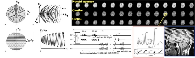
MR image formation is most commonly decribed by linear magnetic gradient fields tracing out trajectories in k-space, followed by image reconstruction via a multidimensional Fourier transform. Spectroscopic imaging then becomes a straightforward extension whereby chemical shift information (used to distinguish different metabolic components) is encoded as an additional k-space axis. One of our ongoing research projects is to investigate of the use of time-varying readout gradients in combination with RF coil arrays to improve spatial coverage, increase spatial resolution, and reduce acquisition times of in vivo studies. Spiral MRSI, as depicted above, is one of our primary tools.

RSL locations

The Richard M. Lucas Center for Medical Imaging
1201 Welch Rd, Stanford CA 94305-5488
Stanford School of Medicine Technology & Innovation Park
3155 Porter Drive, Palo Alto, CA 94304