Overall Goal of the Lab
The Wu Laboratory seeks to identify mechanisms responsible for human heart diseases, the most common cause of death in the U.S. and the biggest contributor to morbidity and mortality worldwide. We believe that by understanding the mechanisms regulating the formation of the heart during fetal development we can then apply these principles to understand the causes of adult heart diseases such as heart attack and heart failure. We currently use laboratory mouse to help us understand the biology of heart formation. We also use human stem cells as a test-tube model to study the process of heart formation and to create new human heart tissues and organs from these stem cells.
Single Cell Multi-Omics and Bioinformatics
Project Description – The Wu Laboratory was among the earliest labs in the country to engage single cell biology using novel single cell tools as they become available. We were interested in the developmental potential of single cardiac progenitor cell into cardiomyocytes, smooth muscle cells, and endothelial cells. We subsequently became interested in developing a developing heart expression atlas in order to identify the cell type, developmental stage, and anatomical location of each single cardiac cells during mouse heart development using a random forest-based computational algorithm that we termed ATLAS-seq. We have more recently acquired the expertise to execute multi-omic technologies based on single cell microfluidic tools developed from 10x Genomics including single nuclear ATAC-seq with RNA-seq, single cell TCR sequencing with feature barcoding of surface protein (i.e. CITE-seq) for immune cell phenotyping, and multiplexing samples from different sources using custom-made antibodies or lipid binding dye (i.e. MULTI-seq) with oligonucleotide tags.
Some of our current projects include:
1. Development of a ML/AI pipeline for the prediction of subtypes and differentiation stage of developing mouse cardiac cells from mesoderm to birth and from in vitro differentiation of human induced pluripotent stem cells (hiPSCs).
2. Understanding the epigenetic/regulatory landscape of the developing cardiac progenitor cells, their progenies, and the cardiac conduction system cells using single cell multi-omic technologies
3. Identification of causal cardiac antigen involved in ICI-mediate myocarditis in cancer patients and in mouse model of PD-1/PD-L1 loss of function using single cell proteomics, transcriptomics, and genomic TCR sequencing
4. Functional analysis of genome wide association study (GWAS)-implicated genes for atrial arrhythmia using data from single cell RNA sequencing and CRISPR-mediated perturbations studies
Recent Accomplishments - Starting in 2013, we incorporated Fluidigm-based multiplex single cell PCR equipment HD Biomark to analyze the differentiation of single cardiac progenitor cells in vitro and found that Nkx2.5 expression marks a subpopulation of committed endocardial precursor cells in the mouse heart (Li et al, Development 2015). Shortly after, we adopted the Fluidigm C1 microfluidic single cell capture platform and profiled >2300 cells from fetal mouse hearts at embryonic day 8.5, 9.5, and 10.5 of development by RNA sequencing (Li et al, Developmental Cell, 2016; Li et al, Development 2019) as well as the developing cardiac conduction system at embryonic day 16.5 of mouse development (Goodyer et al, Circulation Research 2019). In parallel, we contributed to the cardiac cell isolation and analysis work of the Tabula Muris Consortium which led to multiple publications describing transcriptome profile of single cells from 18 organs (>85,000 cells) in the 3 month old mice (Tabula Muris Consortium, Nature 2018) and during the aging process from the neonatal stage to the end of life (Tabula Muris Senis, Nature, 2020; Schaum et al, Nature 2020). Recently, we have begun to address the pathogenic immune response in patients with myocarditis from treatment with immune checkpoint inhibitors (ICIs) for cancer therapy using single cell immunology tools from 10x Genomics. As the analysis of these data is computationally intensive, we have expanded our technology platform to incorporate biocomputational work including machine learning/artificial intelligence (ML/AI) to enhance our understand of the biological meaning from our single cell RNA seq data.
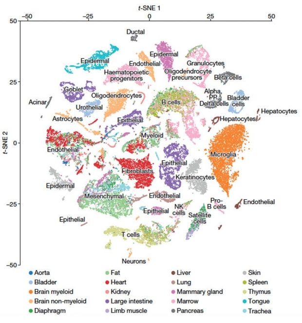
Tabula Muris Consortium
Nature 2018
Cardiovascular Developmental Biology
Project Description – A major focus of the Wu Laboratory is to define the earliest steps in heart formation. We use experimentally-modified mice as our live model to take advantage of a broad range of molecular tools available. The similarity between a mouse heart and a human heart allows us to connect our results directly into finding ways to treat human heart diseases. We seek to understand what genes are responsible for making the heart chamber form in the right way. We are also interested in finding out what disturbances in the normal process of heart formation is responsible for devastating congenital heart diseases that lead to fetal demise or death shortly after birth. We have utilized the most state-of-the-art tools to try to understand the process of normal heart formation and have made significant discoveries in this area of research. Some of the current research project that our ongoing in the laboratory include:
- Identify key genes that regulating commitment of heart precursor cells into cardiac muscle cells and cells of the blood vessels such as smooth muscle cells and endothelial cells.
- Determine, at the single cell level, the key genes that indicate where that cell is localized in the developing heart. This is akin to finding the true “address code” of each single heart cell during heart formation.
Recent Accomplishments – We have isolated the earliest committed heart precursor cell, a master heart stem cell, that can give rise to all major cell types in the developing heart (Wu et al, Cell 2006).
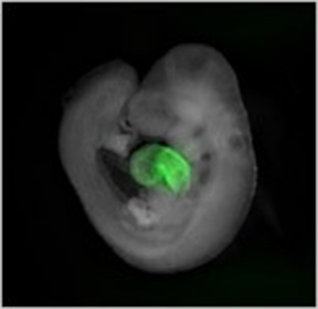
Expression of eGFP Specifically in the Developing Heart Tube. Transgenic mouse embryo at embryonic day 9.5 is directed to express eGFP under a cardiac-specific enhancer from the murine Nkx2.5 locus. Image shown is a composite of eGFP signal (in green) with the body of the embryo (in white).
We have also identified a master regulator of early heart development called Yin Yang 1 that is absolutely required for the existence of heart cells (Gregoire et al, Circ Res 2013). Very recently, we have uncovered a panel of key genes that can be used to identify the precise address code of each individual mouse heart cell during fetal development (Li et al, Dev Cell 2016).
Cardiovascular Tissue Engineering
Project Description – We have embarked on cardiac tissue engineering relatively recently due to the significant promise of this research direction in creating functional cardiac tissue for modeling of heart diseases and for generation a new organ that may be transplantable. By using stem cells that can be turned into cardiac cells, we have brought stem cell biology and tissue engineering together to begin making true functional heart tissue for screening drugs to treat heart diseases and to build new replacement tissues that may one day be used to replace the damaged heart muscle after heart attack. We have actively collaborated with material science engineers, vascular engineers, and mechanical engineers to make new discoveries in this research area. Some of the current research project that our ongoing in the laboratory include:
- Employ 3D bioprinting as a tool to generate full-thickness, vascularized, and functional cardiac tissue that can be perfused readily for maintaining cell survival long-term.
- Evaluate the friendliness of various biomaterials to cardiac muscle cells derived from induced pluripotent stem cells and create 3D scaffold using these biomaterials to support the survival and function of 3D cardiac muscle cells.
Recent Accomplishments – We have successfully partnered with Advanced Solution Life Sciences, Inc. to acquire a BioAssemblybot 3D bioprinter into our research lab and have now used this 3D bioprinter to make various 3D cardiac tissues that can be perfused with nutrients and oxygen for long-term survival. We have evaluated and successfully incorporated stem cell-derived cardiac muscle cells into highly novel biomaterials such as functionalized gelatin and elastin-like peptides and showed that these materials are supportive of cardiac muscle cell function.

Figure 1
The BioAssemblyBot (BAB) 3D bioprinter in the Sean Wu lab at the Stanford University Cardiovascular Institute; World's first 6-axis robotic arm for agile and advanced tissue biofabrication.
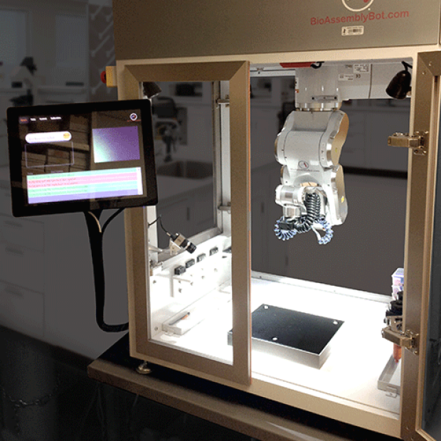
Figure 2
The BioAssemblyBot (BAB) 3D bioprinter in the Sean Wu lab at the Stanford University Cardiovascular Institute. Photos shows the entire setup including the robotic arm, the human-machine interface (HMI), printing stage, video camera, 8 bioink storages, all incorporated inside the printing cabinet.
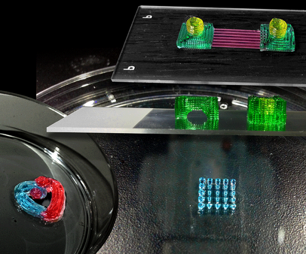
Figure 3
Different printed constructs created by the BAB printer, including the Stanford Cardiovascular Institute (CVI) logo, 3D mesh structure, bridge and embedded cubic constructs, and microchannels printed between two flow chambers. All constructs are printed with pluronic sacrificial bioink.

Figure 4
Snapshot demonstrating bioprinting of a cardiac tube. hiPSC-CMs were suspended in gelatin methacrylated (gelMA) as bioink and printed into a cylindrical construct. The tissue was subsequently cured via exposure to ultraviolet (UV) light (bottom).

Figure 5
Printing the vascular tree network using two different pluronic bioinks, mimicking the multi-layer vessel wall structure.

Figure 6
3D bioprinting of functional liver tissue, mimicking the hexagonal structure of liver tissue lobules. Primary human hepatocytes within the printed honeycomb construct demonstrated a healthy morphology and significant viability-function. The constructs are now being tested in the mouse model.

Figure 7
The BioAssemblyBot (BAB 2.0) 3D bioprinter in the Sean Wu lab at the Stanford University Cardiovascular Institute. The printer is equipped with cooling and heating nozzles, temperature-controlled printing stage, up to 8 printing nozzles (can be used simultaneously), and a state-of-the-art 6-axis robotic arm, enabling deposition of soft cellular bioinks at 50-100 µm resolution.
Cardiovascular Disease Modeling
Project Description – While mouse models are useful for studying the process of heart formation, they are not exactly like the human hearts in various ways. Since we cannot easily obtain human heart tissue, we have chosen to use stem cells as the next best source of material to study human heart formation and disease onset. We focus on a special type of stem cells call induced pluripotent stem cells (iPSCs) that behave exactly like embryonic stem cells but are made from regular human skin or blood cell. These human iPSCs make excellent model of heart formation inside a petri dish in the lab and can be turned into beating heart muscle cells by treating them with special factors.
Beating Cardiomyocytes Derived from In Vitro Differentiated ES Cells. CJ7 ES cells were electroporated with a-MHC-eGFP vector and selected for stably integrated clones that expresses eGFP specifically in beating cardiomyocytes. Video shown represents fluorescence microscopy images of one such ES cell clone that has differentiated for 17 days.
Furthermore, the steps that these iPSCs take to become heart muscle cells replicate exactly the way a human fetus goes through during early development in utero. Some of the current research project that our ongoing in the laboratory include:
- Generating special reagents that allows for isolation of specific heart chamber cells such as ventricular or atrial cells or pacemaker cells.
- Evaluate the molecular defect that occurs when iPSCs made from patients with congenital heart diseases such as Hypoplastic Left Heart Syndrome, Ventricular Septal Defect, or Atrial Septal Defect, or adult heart diseases such as hypertrophic cardiomyopathy, dilated cardiomyopathy, and neutral lipid storage disease undergo differentiation into heart cells.
Recent Accomplishments – We have described the key molecular mechanism of heart muscle cell formation (Galdos et al, Circ Res 2017) as well as the use of human iPSC to model viral myocarditis (Sharma et al, Circ Res 2014). We have also described the generation of human, pig, rat, and mouse iPSCs (Kuppusamy et al, Curr Prot Mol Biol, 2012). Furthermore, we have shown that iPSCs from mouse and human model of neutral lipid storage disease can be use to identify new drugs for therapy (Huang et al, JCI, In Preparation).
Cardiovascular Regenerative Biology
Project Description – Ultimately, our work in developmental biology and tissue engineering seek to identify the most effective way to treat damage hearts. The regenerative potentials of stem cells is unlimited but requires careful guidance when given to a patient with heart disease. Many efforts that have failed in the past is due to the lack of understanding of what stem cells are capable of doing to treat damaged heart. We have studied the role of stem cells in a fetal heart injury and recovery model (Sturzu et al, Circulation 2015) and have addressed the challenges that must be overcome in order to move the field forward (Wu et al, Cell 2008). We are currently seeking to find new cell types that may be useful for repairing damages to the conduction system (i.e. the electrical network) in the heart using human iPSC-derived cells. Some of the current research project that our ongoing in the laboratory include:
- Identifying genes that are unique to conduction system cells such as purkinje fiber such that these cells can be isolated from differentiating human iPSCs for therapy and investigation.
- Generation of functional organ by chimera complementation of human or animal stem cells into early animal fetuses that are incapable of making a normal heart on its own. This allows for the creation of a fully normal/functioning heart that is composed entirely from the stem cells that were introduced.
Recent Accomplishments – We have recently published a major article on the need for better understanding of heart development to improve heart regeneration (Galdos et al, Circ Res 2017). We have also discussed the various stem cell treatments that have been tried in patients with heart attack (Sturzu et al, Circ Res, 2011). We showed that the fetal mouse heart is capable of losing up to 60% of its heart cells and is still able to recover itself to a normal functioning heart (Sturzu et al, Circulation, 2015). We also showed that one can make a normal mouse heart that is derived from stem cells introduced into a mouse fetus during early development (Sturzu et al, In Preparation, 2017). We also found that a rare population of heart stem cell is able to mediate the repair a newborn heart (Chen et al, Cardiovasc Res, 2014).