ISMRM Honors 2016
RSL Celebrates Award Winning Research
Summa Cum Laude
Michael Marx1, Pejman Ghanouni1, and Kim Butts Pauly1
1Radiology, Stanford University, Stanford, CA, United States
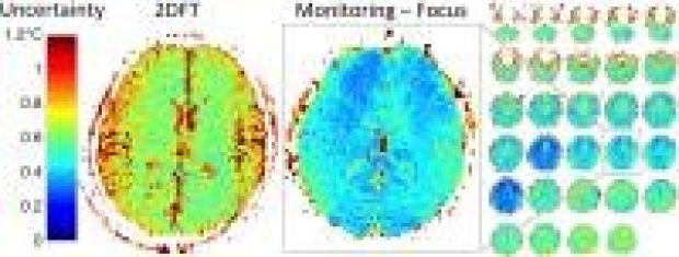
Multi-slice thermometry was developed that overcomes several limitations of single-slice 2DFT thermometry in MR-guided focused ultrasound brain treatment. Using multiple-echo spiral imaging provides much greater imaging performance, which was applied to improved focal spot localization and to improved ablation monitoring. High-resolution higher-precision multi-slice focal spot localization can shorten treatment time and improve patient safety. High-speed high-precision focal spot monitoring, combined with full-brain monitoring and 3-dimensional focal spot characterization during ablations can improve treatment guidance and feedback while also improving patient safety. The new sequences were validated both in vivo and in a phantom within a clinical transducer.
Qiyuan Tian1,2, Max Wintermark2, Kim Butts Pauly2, Diane Huss3, W. Jeffrey Elias4, and Jennifer A. McNab2
1Electrical Engineering, Stanford University, Stanford, CA, United States, 2Radiology, Stanford University, Stanford, CA, United States, 3Physical Therapy, University of Virginia, Charlottesville, VA, United States,4Neurosurgery, University of Virginia, Charlottesville, VA, United States
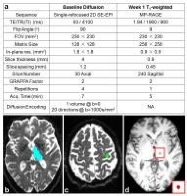
We retrospectively studied 13 essential tremor patients treated with MRI-guided focused ultrasound. The purpose was to demonstrate the value of using diffusion MRI tractography to help localize the ventral intermediate (Vim) nucleus of the thalamus (the treatment target). Tractography between the thalamus and hand-knob region of the motor cortex was consistent from subject-to-subject and followed the expected anatomy. The thalamic voxels with high tractography streamline counts qualitatively matched the location of Vim as depicted on the Schaltenbrand-Wahren Atlas. A trend was found towards better treatment outcome scores with higher pre-treatment probabilistic tractography streamline counts within the visualized MRgFUS treatment-induced lesion.

Christian Federau1, Maged Goubran1, Jason Su1, Jaimie Henderson1, Veronika Santini1, Casey Harrison Halpern1, Brian Rutt1, Kim Butts Pauly1, and Pejman Ghanouni1
1Stanford University, Stanford, CA, United States
Transcranial MR-guided high-intensity focused ultrasound ablation of the ventral division of the ventral lateral posterior thalamic nucleus (VLpv) is a promising, minimally invasive treatment method for essential tremor. We report our initial clinical experience in 11 patients, and correlate clinical outcome with lesion size, location, and thermal dose during treatment. We found a correlation between clinical outcome at 1 year follow-up and lesion size (r = 0.73), as well as thermal dose in the VLpv (r = 0.65).
Nuisance Regression of High-frequency FMRI Data: De-noising Can Be Noisy
Jingyuan E. Chen1,2, Hesamoddin Jahanian2, and Gary H. Glover1,2
1Electrical Engineering, Stanford University, Stanford, CA, United States, 2Radiology, Stanford University, Stanford, CA, United States
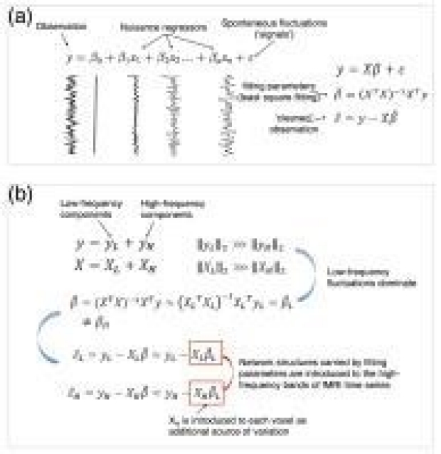
A growing number of studies using fast sampling have demonstrated the persistence of functional connectivity (FC) in resting state (RS) networks beyond the conventional 0.1 Hz. However, some RS studies have reported frequencies (e.g., up to 5 Hz) not easily supported by canonical hemodynamic response functions. Here, we investigated the influence of a common preprocessing step – whole-band (the entire frequency band resolved by a short TR) linear nuisance regression (LNR) – on RSFC. We demonstrated via both simulation and real data that LNR can introduce network structures in HF bands, which may largely account for the observations of HF-RSFC.
Dynamic analysis of [18F]-sodium fluoride uptake in knee osteoarthritis with PET-MRI
Audrey P Fan1, Feliks Kogan1, Aleema Patel1, Edwin HG Oei2, Andrew Quon1, and Garry E Gold1
1Radiology, Stanford University, Stanford, CA, United States, 2Erasmus MC: University Medical Center Rotterdam, Rotterdam, Netherlands
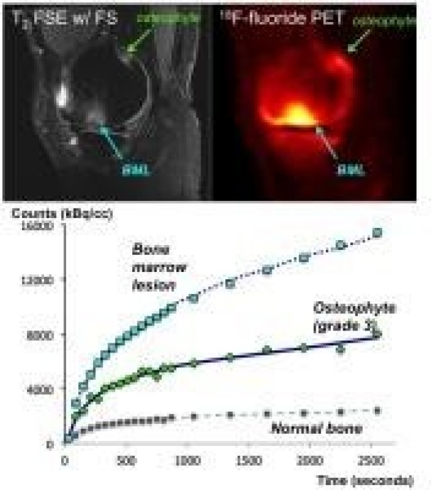
This study investigates dynamic uptake of [18F]-fluoride in bone marrow lesions (BMLs) and osteophytes observed on MRI of patients with knee osteoarthritis. Through kinetic modeling, we characterized rate constants of bone metabolism in bone pathology relative to healthy bone. BMLs and higher-grade osteophytes showed higher total bone metabolism Ki (P < 0.01) and higher bone mineralization rate k3 (P < 0.01) relative to grade 1 osteophytes and normal bone. While a similar trend was observed for blood flow, the differences from normal tissue were subtler suggests that rate of mineralization k3 and not blood flow is a key driver of [18F]-fluoride accumulation in OA lesions. These new physiological parameters may help differentiate between different grades of OA lesions or identify which lesions are active parts of the disease process.

Stroke Volume Evolution Following Endovascular Therapy on DWI and FLAIR
Christian Federau1, Soren Christensen1, Michael Mlynash1, Jenny Tsai1, Sun Kim1, Greg Zaharchuk1, Matus Straka1, Nishant Mishra1, Maarten Lansberg1, and Greg Albers1
1Stanford University, Stanford, CA, United States
We studied the evolution of the infarct volume between an early post-revascularization scan (within 24 h of symptom onset) and day 5 in patients of the CRISP and DEFUSE 2 cohort studies. On the early post-revascularization scan, FLAIR lesions were smaller compared to DWI, but were larger at day 5. The early post-revascularization stroke volume on DWI, compared to FLAIR, was closer, and c

Quantification and Artifact Reduction from Simple Modeling of DESS Signals
Bragi Sveinsson1, Garry Gold1, and Brian Hargreaves1
1Stanford University, Stanford, CA, United States
The double-echo in steady-state (DESS) sequence offers both 3D anatomical imaging and 3D quantitative mapping (SNR-efficient 3D maps of T2 and apparent diffusion coefficent) in various applications, such as breast imaging or knee cartilage imaging. The complicated signal behavior remains a challenge for quantitative imaging, and strong spoiling can lead to motion artifacts. Here, we introduce simplified methods for modeling DESS signals, enabling more accurate T2 measurements and better motion artifact reduction.
Magna Cum Laude

Observation of in vivo lactate metabolism in skeletal muscle using hyperpolarized 13C MRS
JAE MO PARK1, Sonal Josan1, Dirk Mayer2, Ralph E Hurd3, Youngran Chung4, David Bendahan5, Daniel M Spielman1, and Thomas Jue4
1Radiology, Stanford University, Stanford, CA, United States, 2Diagnostic Radiology and Nuclear Medicine, University of Maryland, Baltimore, MD, United States, 3Applied Sciences Laboratory, GE Healthcare, Menlo Park, CA, United States, 4Biochemistry and Molecular Medicine, University of California - Davis, Davis, CA, United States, 5Centre de Resonance Magnetique Biologique et Medicale, Aix-Marseille University, Marseille, France
The present study reports the use of hyperpolarized [1-13C]lactate and [2-13C]pyruvate to measure the rapid pyruvate and lactate kinetics in rat skeletal muscle. The results provide support for a critical underpinning of both the glycogen shunt model and the intracellular lactate shuttle hypothesis, and cautions against an overly simplistic view of glycolytic end products as merely hypoxia biomarkers.
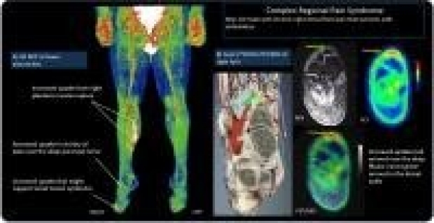
PET/MRI Of Patients With Chronic Pain Alters Management: Early Experience.
Daehyun Yoon1, Deepak Behera1, Dawn Holley1, Pamela Gallant1, Ma Agnes Martinez Ith2, Ian Carroll3, Matthew Smuck2, Brian Hargreaves1, and Sandip Biswal1
1Radiology, Stanford University, Palo Alto, CA, United States, 2Orthopaedic Surgery, Stanford University, Palo Alto, CA, United States, 3Anesthesia, Stanford University, Palo Alto, CA, United States
The chronic pain sufferer is currently faced with a lack of objective tools to identify the source of their pain. Increased inflammation of the nervous system, vessels, muscles, and other tissues in chronic pain sufferers and [18F]fluorodeoxyglucose positron emission tomography/magnetic resonance imaging ([18F]FDG PET/MRI) has emerged as a sensitive clinical tool to identify increased inflammation. We plan to develop clinical [18F]FDG PET/MRI method to more accurately localize sites of hypermetabolic foci as it relates to pain generators. Early clinical results suggest that [18F]FDG PET/MRI can identify abnormalities in chronic pain patients and can immediately affect their management.

Clinically Viable Diffusion-Weighted Imaging Near Metal using 2D-MSI PROPELLER DUO
Suryanarayanan Sivaram Kaushik1, Ajeet Gaddipati2, Brian Hargreaves3, Dawei Gui4, Robert Peters2, Tugan Muftuler5, and Kevin Koch1
1Radiology, Medical College of Wisconsin, Milwaukee, WI, United States, 2GE Healthcare, Waukesha, WI, United States, 3Radiology, Stanford University, Stanford, CA, United States, 4GE Healthcare, Waukesh, WI, United States, 5Neurosurgery, Medical College of Wisconsin, Milwaukee, WI, United States
While FSE-based multi-spectral imaging (MSI) sequences help overcome the artifacts caused by metallic hardware, diffusion-weighted imaging remains a challenge. The non-CPMG artifacts caused by adding diffusion lobes to an FSE train can be mitigated by modulating the phase of the refocusing pulses. Another solution involves splitting the contribution made by the spin and stimulated echoes (DUO acquisition). Here, we combine a 2D version of MSI with a PROPELLER-DUO sequence to obtain clinically-feasible, artifact-minimized, diffusion-weighted images in subjects that have cancerous lesions in close proximity to metallic hardware.

In vivo 7T Quantitative Susceptibility Mapping of Cortical Lesions in Multiple Sclerosis
Wei Bian1, Eric Tranvinh1, Thomas Tourdias2, May Han3, Tian Liu4, Yi Wang4, Brian Rutt1, and Michael Zeineh1
1Department of Radiology, Stanford University, Stanford, CA, United States, 2Service de NeuroImagerie Diagnostique et Thérapeutique, CHU de Bordeaux, Bordeaux, France, 3Department of Neurology, Stanford University, Stanford, CA, United States, 4Department of Radiology, Weill Medical College of Cornell University, New York, NY, United States
Magnetic susceptibility measured with quantitative susceptibility mapping (QSM) has been proposed as a biomarker for inflammation in multiple sclerosis (MS) white matter (WM) lesions. However, a detailed in vivo characterization of cortical lesions has not been performed. In this study, the susceptibility in both cortical and WM lesions relative to adjacent normal-appearing parenchyma was measured and compared for 14 MS patients using QSM at 7T. Our results showed that relative susceptibility was negative for cortical lesions but positive for WM lesions. The opposite pattern of relative susceptibility suggests that iron loss dominates the susceptibility contrast in cortical lesions.
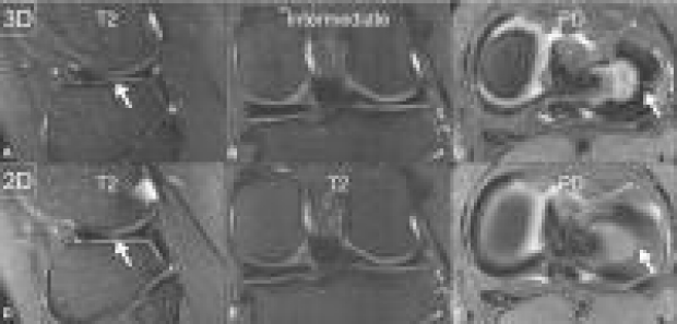
Fast single sequence comprehensive 4D pediatric knee MRI with T2 Shuffling
Shanshan Bao1, Jonathan I. Tamir2, Umar Tariq3, Martin Uecker4, Peng Lai5, Weitian Chen5, Michael Lustig2, and Shreyas S. Vasanawala1
1Radiology, Stanford University, Stanford, CA, United States, 2University of California, Berkeley, Berkeley, CA, United States, 3Geisinger Medical Center, Danville, PA, United States, 4University Medical Center Göttingen, Göttingen, Germany, 5GE Healthcare, Menlo Park, CA, United States
Clinical application of volumetric joint MR imaging has been hampered by blurring due to T2 decay. A redesigned volumetric fast spin-echo acquisition technique termed T2 shuffling corrects for T2 decay and yields effectively a four-dimensional reconstruction with varying degrees of T2 weighting. Our work assesses the clinical application of T2 shuffling for pediatric knee MRI. Our results show that T2 shuffling has the potential to suffice as a single sequence MR examination. This is especially relevant for pediatric imaging where streamlined protocols greatly improve clinical operations and patient experience.