A new cell imaging technology helps researchers investigate biological processes
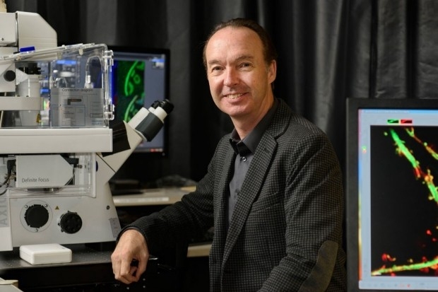
Jon Mulholland, Director of the Stanford Cell Sciences Imaging Facility (CSIF)
Photo: Steve Fisch
Stanford has introduced a new investigative tool to help researchers visualize and study biological processes through the use of microscopy and molecular probe technologies. By marking molecules with multicolored tags, the inner workings of cells can be transformed into observable light shows, illuminating previously invisible processes and interactions in the context of complex tissues or organs, such as the way microbes invade cells or how immune system cells destroy tumors.
The Stanford Cell Sciences Imaging Facility, a core research service center in the School of Medicine, is located in the Beckman Center and the Shriram Center, trains researchers to use light and electron microscopes, prepare biological samples and interpret resulting data. The facility recently launched a new imaging service that uses the Akoya Inc’s CODEX system, a novel technology that enables researchers to color-code and track up to 50 different molecules in a single tissue sample, ten times the number available with conventional technologies.
In traditional fluorescence experiments, researchers are limited to seeing about five distinct proteins at once, each labeled with a different color fluorescent tag. CODEX expands the number of molecules that can be simultaneously imaged by combining red, green and blue fluorescent molecules with a library of unique DNA oligo barcodes that are complimentary to those attached to the tissue- or protein-specific antibodies. For understanding cancer cells—which involve dozens of molecular players interacting in many ways, being able to see more proteins and their subcellular organization simultaneously is a huge boon to research.
The core of the CODEX system is a small microfluidics device engineered to fit on a standard fluorescence microscope platform. The tissue sample remains stationary during sequential, probe localization imaging rounds, so that each picture can be overlaid into one high resolution, multiplexed image. The multiplexed sequencing capability of the CODEX allows large numbers of probes to be pooled and imaged during a single run. Akoya’s advanced software then assists researchers in identifying classes and relationships between cells.
“Since you can see where various phenotypes or classes of cells are simultaneously positioned relative to each other, you can start to make hypotheses about interactions between those cells, and whole networks of cells,” says Peter Jackson, PhD, professor of microbiology and immunology, who has worked with CODEX in his own lab.
As an example, this technology provides researchers with insights into what molecular markers on cells might be useful to target with anti-cancer drugs. It also lets them compare how different classes of cancer cells change — or disappear — after different treatments, which can shed light on why some drugs work better for some patients than others.
The CODEX system was originally developed by Garry Nolan, PhD, professor of microbiology and immunology, and commercialized by Akoya Biosciences. The instrumentation and staff are supported by the Senior Associate Dean of Research’s office in the School of Medicine, by the Beckman Center for Molecular and Genetic Medicine, and by the Cancer Institute.
To request training or access equipment at the Stanford Cell Sciences Imaging Facility, go to https://microscopy.stanford.edu or contact the director, Jon Mulholland, at jwm@stanford.edu.