Retinal Prosthesis
The leading cause of blindness in developed countries is Age-related Macular Degeneration (AMD), which results in severe and irreversible loss of vision for over two million Americans. The disease causes deterioration of the photoreceptor cells in the central part of the retina, and currently, there is no cure. The retinal neurons that receive signals from the photoreceptors, however, are mostly preserved even several years after the onset of blindness. A possible therapeutic solution is to communicate with these neurons directly to restore the visual pathway.
It has been already demonstrated by several groups that electric stimulation of the retina can produce perception of light ("phosphenes") in patients suffering from retinal degeneration. Current devices for retinal stimulation involve a very small number of electrodes, while several thousands of pixels are required for functional restoration of sight. Number of electrodes in the array is limited by such physical factors as heating of the retina, cross-talk between neighboring electrodes and electrochemistry at the electrode-liquid interface. All these factors are strongly dependant on the distance between the electrodes and the target cells. For the pixel density geometrically corresponding to visual acuity of 20/400 (the level of "legal blindness") the cells should not be farther away from electrodes than 30 micrometers, and for visual acuity of 20/80 this distance should not exceed 7 micrometers. Such stringent requirements of proximity between thousands of electrodes and their target cells preclude application of a flat array of electrodes for high resolution retinal stimulation on either epiretinal or subretinal sides.
We work on high resolution retinal prosthesis using several different approaches to (a) bringing neural cells into proximity of stimulation sites, (b) mechanisms of stimulation, and (c) transfer of information and power to the implant.
Bringing cells to electrodes
Directed neural growth
One way of bringing the target cells to stimulation sites is based on application of the soft lithographic techniques, such as microcontact printing, for directing nerve fiber growth to individual microelectrodes to achieve single cell stimulation. We have found that micropatterning techniques significantly reduce the current and power requirements necessary for neuronal stimulation. We have also fabricated microelectrode arrays with various geometries at the Stanford Nanofabrication Facility to identify and investigate the governing electrical parameters for the stimulation of micropatterned neurons.

Brightfield image of micropatterned retinal ganglion cells (RGCs)
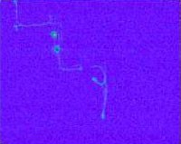
Fluorescence image of micropatterned RGC electrical stimulation
Migration of Retinal Cells
Another approach to bringing cells into proximity of the stimulation sites is based on a fascinating effect of retinal migration. Recently we discovered that retinal cells rapidly (within 48-72 hours) migrate into a perforated sub-retinal implant while preserving their axonal connections to the retina above the implant. Within the pores in the implant stimulating electrodes can be positioned, and this way an intimate proximity between electrodes and target cells is achieved automatically along the whole surface of the implant.
Bringing electrodes to cells
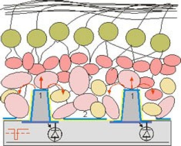
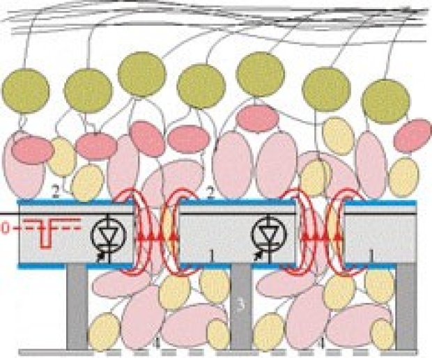
Effect of cellular migration can also be utilized with an implant having an array of thin protruding electrodes insulated at their sides and exposed at the tops. When positioned under the retina, the cells migrate into the spaces between the pillars thus assuring penetration of the electrodes into the retina without high pressure and associated risk of mechanical injury. The depth of penetration is determined by the length of the electrodes. The approaches based on pores and on protruding electrodes are complimentary: in the first case the actively migrating cells penetrating into the pores will be stimulated. In the second case the actively migrating cells move towards the bottom of an implant, while the electrodes approach the target cells which did not migrate.
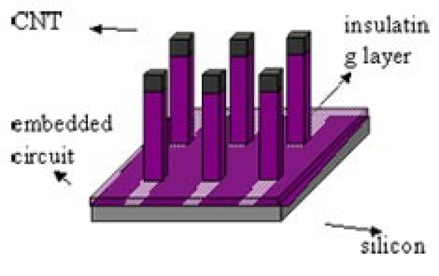
Penetrating carbon nanotubes
Traditionally the retinal prosthetic electrodes are mage of metals (Pt, Au) and metal derivatives (IrOx, TiN). Common problems include biocompatibility and electrochemical stability. We are trying to improve these issues by developing a novel electro-neural interface using vertically self-aligned carbon nanotube (CNT) bundles as flexible, protruding microelectrodes. CNT bundles are robust, flexible, conductive and appear to be biocompatible, they also have superior electrochemical properties. All these indicate that the CNT protruding electrodes may be able to provide a safer solution for long-term retinal stimulation and implantation. They could also act as recording units to sense electrical and chemical activities in neural systems for fundamental neuroscience research.

Carbon nanotubes are graphite sheets wrapped seamlessly into cylindrical tubes.
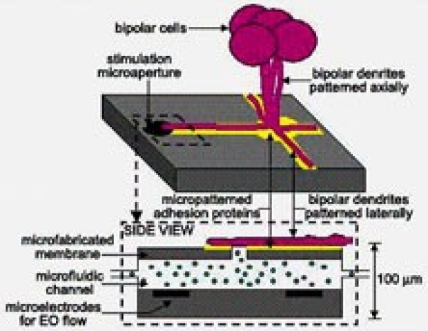
Actuated neurotransmitter-based stimulation
There is great concern, however, over the immune response to chronic electrical stimulation as well as the ability of such a method to harness the biological specificity of retinal circuitry (such as the ON and OFF pathways that provide spatial contrast). The primary means of nerve cell stimulation physiologically is through synaptic transmission, a process in which the localized delivery of neurotransmitters occurs between cell junctions. For this reason, we are also investigating the feasibility of a retinal interface based on localized chemical stimulation. Using advances that have been made in the rapidly developing field of microfluidics, soft materials engineering techniques are being used to microfabricate a neurotransmitter delivery device that conforms to the curvature of the retina. Cell micropatterning in three dimensions will be used to direct individual retinal neurons to stimulation sites on the retinal interface. Photodiodes will be incorporated into the microfluidic network to transduce a light signal to actuate fluidic release using either electroosmotic flow or electrowetting. The integration of directed neurite growth and actuated neurotransmitter stimulation at a high resolution would represent a novel approach in the growing field of neural prostheses. The development of such an interface may provide the physiologic approach that is necessary not only to treat retinal degeneration, but a variety of neurological disorders as well.

Multiple stimulation sites
Single stimulation site
Optoelectronic System Design
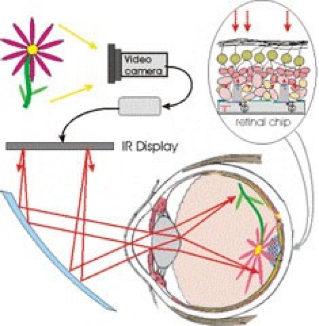
We are developing an optical system for a parallel delivery of information and energy to tens of thousands of pixels in the implant. This system permits normal eye scanning to observe a large field of view, as opposed to "hard wiring" of a video camera to the retinal stimulating array using a single emitter-receiver link. The image from a goggles-mounted video camera is processed in a portable microcomputer and then projected with a pulsed IR LED-LCD array onto the retina. The retinal implant has an array of powered photodiodes converting pulses of light into pulsed electrical current using a power from an intraocular photovoltaic battery.
Optoelectronic System Design
Principal Investigators:
Daniel Palanker, Ph.D.
Michael Marmor, MD.
Mark Blumenkranz, MD.
Students and staff members:
Roopa Dalal, MSc.
Collaborators:
Stacey Bent, PhD. (Chem. Eng.)
James Harris, PhD. (Elec. Eng.)