Mutations in immune cells prevented their natural death in roughly half of the cell samples from patients with the incurable cancer, and suggest drug targets for the disease.
August 10, 2015 - By Kim Smuga-Otto
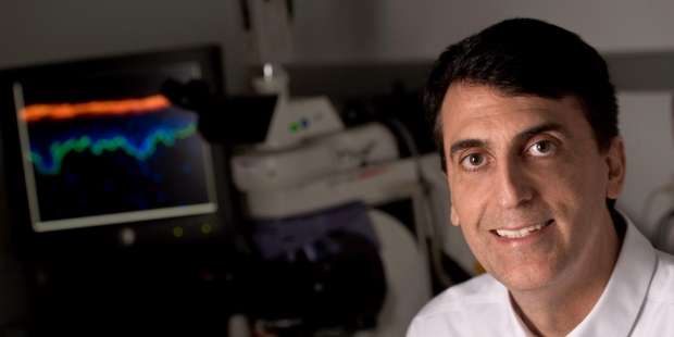
Paul Khavari and his colleagues have identified mutations that can cause a rare immune cell cancer.
Steve Fisch
Researchers at the Stanford University School of Medicine have identified a group of mutations responsible for many cases of a rare immune cell cancer called cutaneous T-cell lymphoma. Identifying the mutations could tip off clinicians to effective treatments for the currently incurable condition.
The mutations are found in 15 genes for proteins that work together in a T-cell-survival mechanism. When mutations prevent the mechanism from switching off, the T cells don’t die when they should and actually keep multiplying.
The findings were published online Aug. 10 in Nature Genetics.
Cutaneous T-cell lymphoma — which manifests as rashes, skin tumors and leukemia — doesn’t respond well to traditional chemotherapy, although a technique known as total skin electron radiation that was developed at Stanford can keep the skin disease at bay. Additionally, a new stem cell transplant therapy, also from Stanford, shows promise for long-term remission for those patients with advanced, high-risk disease.
The newly identified cancer role of the proteins involved in the cell-survival mechanism suggests new strategies for fighting the disease: Researchers can look for drugs to counter the malfunctioning proteins resulting from the mutations.
“We can now design drug trials in a smart, evidence-based way that is specific to the patient,” said Youn Kim, MD, professor of dermatology and one of the paper’s authors.
We can now design drug trials in a smart, evidence-based way that is specific to the patient.
But before doctors can prescribe drugs targeted to specific proteins, they’ll need to know which, if any, mutations in the cell-survival pathway their patients’ malignant T cells carry. In 60 percent of these cancers sequenced by the researchers, such mutations were absent.
“It really highlights that the future of many types of cancer treatment is going to be: first, know the cancer by sequencing it, and then tailor the therapy specifically,” said senior author Paul Khavari, MD, PhD, professor and chair of dermatology.
Lead authorship is shared by graduate student Aparna Bhaduri, postdoctoral scholar Eon Rios, MD, PhD, and former postdoctoral scholar Alexander Ungewickell, MD, PhD.
Only about 3,000 patients are diagnosed with cutaneous T-cell lymphoma each year in the United States. The most common form of the disease, mycosis fungoides, resembles common skin rashes — itchy, scaly, red skin eruptions that can cover the whole body. Although it manifests in the skin, the actual cancer cells are roaming immune cells called T cells that are programmed to defend the body from invaders in healthy individuals. In mycosis fungoides, these T cells remain in the skin and multiply excessively, resulting in rashes. In another stage, called Sézary syndrome, the abnormal T cells circulate in the bloodstream, spreading the cancer throughout the body.
Locked into always-on state
Khavari, the Carl J. Herzog Professor in Dermatology in the School of Medicine, likens skin T cells to patrolling sentries, rotating on and off duty. At the end of their shift, the cell-survival mechanism shuts down and, with no signal, the T cells leave or die. The mutations Khavari’s team found prevent the pathway from turning off, causing T cells to pile up in the skin or circulate through the bloodstream. “More and more sentries keep showing up for duty,” he said. “It’s out of control.”

Youn Kim
Because of Kim’s long commitment to treating mycosis fungoides and Sézary syndrome at Stanford, Khavari and his team were able to recruit 91 patients to sequence specific regions of the T cells’ DNA they suspected might be modified in the cancer. From each patient, they collected both cancerous and healthy cells to compare their DNA. This way, they were able to discount any inherent mutations present in the patient before cancer. They identified 170 genes with mutations that could be related to the cancer.
In four of the patients examined, the researchers identified a mutation that replaced a specific amino acid in the tumor necrosis factor receptor 2, a protein embedded in the cell’s membrane that receives signals from outside the cell. The mutation locked the receptor into an always-on state, preventing the cell-survival pathway from shutting down. Previous independent clinical studies found patients with increased TNFR2 protein in their bloodstream had more aggressive forms of the cancer that were more likely to return quickly after treatment. This led Khavari’s team to look at the other patients’ DNA to see if duplications could account for both the elevated levels in the blood and increased signaling to activate the cell-survival pathway. They found that 10 of the patients had multiple copies of the TNFR2 gene.
The researchers confirmed the biological role of TNFR2 by growing cells in the lab with either the amino acid mutation or the duplicate TNFR2 genes and showing the cell-survival pathway to be more active than normal cells.
‘Smoking gun’
Although only 5 percent of the cancers had the TNFR2 mutation, the fact that it was the exact same mutation was a “smoking gun,” according to Khavari, implicating the cell-survival mechanism’s role in driving certain cutaneous T-cell lymphomas. While over half of the patients with the disease did not have these gene changes, identifying those who do presents new options for treating them.
Another one of the mutations caused a receptor that normally signals the cell-survival pathway to stop to instead activate it further and encourage cell proliferation. This receptor, CTLA4, has been identified in skin cancers, and an antibody that turns off the receptor has been approved as the drug ipilimumab to treat advanced melanoma. But before administering the drug, or less toxic alternatives, a physician would need to know if the patient had the mutated receptor; otherwise ipilimumab would have the opposite effect, deactivate a healthy protein and make the cancer worse.
University of California-San Francisco melanoma specialist Susana Ortiz-Urda, MD, PhD, who was not involved with the study, called the work groundbreaking and said she was impressed that the researchers were able to gather so many patients to identify the rare mutations. Ortiz-Urda, who co-directs the UCSF Melanoma Center, said she thought the next step was “putting the paper to work in a clinical setting” to see if patients with different mutations would respond to different drug treatments.
Before we had this data, it was trial and error — we were totally blind.
Kim, the Joanne and Peter Haas Jr. Professor in Cutaneous Lymphoma Research who directs Stanford’s Multidisciplinary Cutaneous Lymphoma Program, plans to do just that, using the individual patients’ cancer cell genetic sequences to design combinations of drugs that would hit multiple defective proteins to completely shut down the cell-survival mechanism. Khavari’s lab will be working to incorporate the mutations they identified into the DNA of living mice. This will allow them to study the mutated genes’ effects, and the actions of new drugs on those genes, directly.
“Before we had this data, it was trial and error — we were totally blind,” said Kim. “We’re finally taking the blindfolds off.”
Other Stanford-affiliated authors are postdoctoral scholars Carolyn Lee, MD, PhD, Ashley Zehnder, DVM, PhD, Jason Reuter, PhD, and Mahkam Tavallaee, PhD; research assistant Angela Mah; Michael Snyder, PhD, professor of genetics; Robert Ohgami, MD, PhD, clinical instructor of pathology; Dita Gratzinger, MD, PhD, assistant professor of pathology; and flow cytometrist Randall Armstrong.
This study was funded by the National Institutes of Health (grants R01CA142635, F32CA168091, ASHRTAF and F310CA180408), the Office of Research and Development of the U.S. Department of Veterans Affairs, the Dermatology Foundation, the Haas Family Foundation, and the Drs. Martin and Dorothy Spatz Charitable Foundation.
Information about Stanford’s Department of Dermatology, which also supported the work, is available at http://dermatology.stanford.edu.
About Stanford Medicine
Stanford Medicine is an integrated academic health system comprising the Stanford School of Medicine and adult and pediatric health care delivery systems. Together, they harness the full potential of biomedicine through collaborative research, education and clinical care for patients. For more information, please visit med.stanford.edu.

