New research involving people diagnosed with Lou Gehrig’s disease sheds light on how individual neurons control muscle movement in humans — and could help in the development of better brain-controlled prosthetic devices.
June 23, 2015 - By Barbara Feder Ostrov
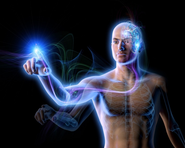
Studying the brain activity of two patients with Lou Gehrig's disease has given researchers insight into how neurons control muscle movement.
Oliver Burston
Stanford University researchers studying how the brain controls movement in people with paralysis, related to their diagnosis of Lou Gehrig’s disease, have found that groups of neurons work together, firing in complex rhythms to signal muscles about when and where to move.
“We hope to apply these findings to create prosthetic devices, such as robotic arms, that better understand and respond to a person’s thoughts,” said Jaimie Henderson, MD, professor of neurosurgery.
A paper describing the study was published online June 23 in eLife. Henderson, who holds the John and Jene Blume-Robert and Ruth Halperin Professorship, and Krishna Shenoy, PhD, professor of electrical engineering and a Howard Hughes Medical Institute investigator, share senior authorship of the paper. The lead author is postdoctoral scholar Chethan Pandarinath, PhD.
The study builds on groundbreaking Stanford animal research that fundamentally has changed how scientists think about how motor cortical neurons work to control movements. “The earlier research with animals showed that many of the firing patterns that seem so confusing when we look at individual neurons become clear when we look at large groups of neurons together as a dynamical system,” Pandarinath said.
Previously, researchers had two theories about how neurons in the motor cortex might control movement: One was that these neurons fired in patterns that represent more abstract commands, such as “move your arm to the right,” and then neurons in different brain areas would translate those instructions to guide the muscle contractions that make the arm move; the other was that the motor cortex neurons would actually send directions to the arm muscles, telling them how to contract.

Krishna Shenoy
But in a 2012 Nature paper, Shenoy and his colleagues reported finding that much more is going on: Motor cortical neurons work as part of an interconnected circuit — a so-called dynamical system — to create rhythmic patterns of neural activity. As these rhythmic patterns are sent to the arm, they drive muscle contractions, causing the arm to move.
“What we discovered in our preclinical work is evidence of how groups of neurons coordinate and cooperate with each other in a very particular way that gives us deeper insight into how the brain is controlling the arm,” Shenoy said.
He and his colleagues wanted to know whether neurons fired similarly in humans.
Recording human brain activity
To conduct the study, the researchers recorded motor cortical brain activity of two research participants with the degenerative neurological condition called amyotrophic lateral sclerosis, or ALS. The condition, which also is known as Lou Gehrig’s disease, damages neurons and causes patients to lose control over their muscles.
The participants, a 51-year-old woman who retained some movement in her fingers and wrists, and a 54-year-old man who could still move one of his index fingers slightly, are participants in the BrainGate2 trial, which is testing a neural interface system allowing thoughts to control computer cursors, robotic arms and other assistive devices.

Jaimie Henderson
These participants had electrode arrays implanted in their brains’ motor cortex for the trial. That allowed researchers to record electrical brain activity from individual neurons while the participants moved or tried to move their fingers and wrists, which were equipped with sensors to record physical movement. Typically, such mapping in humans can only occur during brain surgery.
The participants’ implants provided an “opportunity to ask important scientific questions,” Shenoy said. The researchers found that the ALS patients’ neurons worked very similarly to the preclinical research findings.
Researchers now plan to use their data to improve the algorithms that translate neural activity in the form of electrical impulses into control signals that can guide a robotic arm or a computer cursor.
Other Stanford co-authors of the paper are former research associate Vikash Gilja, PhD; research assistant Christine Blabe; and postdoctoral scholar Paul Nuyujukian, MD, PhD.
The study was funded by the Stanford Institute for Neuro-Innovation and Translational Neuroscience, Stanford BioX/NeuroVentures, the Stanford Office of Postdoctoral Affairs, the Garlick Foundation, the Reeve Foundation, the Craig H. Neilsen Foundation, the National Institutes of Health (grants R01DC009899, N01HD53403 and N01HD10018), the Department of Veterans Affairs and the MGH-Deane Institute for Integrated Research on Atrial Fibrillation and Stroke.
The Department of Neurosurgery, Department of Neurology and Neurological Sciences and Department of Electrical Engineering also supported the work. Information about these departments is available at http://neurosurgery.stanford.edu, http://neurology.stanford.edu and http://ee.stanford.edu, respectively.
About Stanford Medicine
Stanford Medicine is an integrated academic health system comprising the Stanford School of Medicine and adult and pediatric health care delivery systems. Together, they harness the full potential of biomedicine through collaborative research, education and clinical care for patients. For more information, please visit med.stanford.edu.

