A new medical school course brings students to the Cantor Arts Center and Anderson Collection to practice close observation of art, and then learn how to translate those skills to a clinical setting.
March 6, 2015 - By Jacqueline Genovese
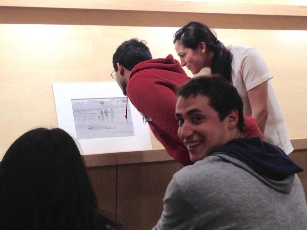
Medical students Sam Cartmell (facing camera) and Abhinav Golla (in red sweatshirt) look at Robert Frank’s Car Accident — U.S. 66, Between Winslow and Flagstaff, Arizona at the Cantor Arts Center, with Sarah Naftalis (far right), a graduate student in art history.
Audrey Shafer
The scene: A group of medical students huddled around the iconic Robert Frank photograph, Car Accident — U.S. 66, Between Winslow and Flagstaff, Arizona, at Stanford’s Cantor Center for the Visual Arts.
Sarah Naftalis, who’s studying for a PhD in art history at Stanford, led the students through an exercise: She asked them what they saw as she gestured to the photograph, which shows four people standing in a field and looking at blanket that presumably covers a corpse, or corpses, on the ground. Several students noted the people, the odd lumpiness of the blanket and the reduced horizon.
Sam Cartmell, one of the medical students, said, “Well, there may be more than four people,” and pointed to an odd contour at the shoulder of the lone woman in the photograph. The observation sparked a lively debate, as his fellow students took turns looking closely at the work, seeking to discern what Cartmell said he’d noticed.
This moment, said Naftalis, illustrates the “productive ambiguities of art,” as well as the benefit of engaged, close looking without “rushing to assign meaning to what we see.”
Welcome to “The Art of Observation: Enhancing Clinical Skills through Visual Analysis,” a new medical school course supported by the Bioethics and Medical Humanities Scholarly Concentration. The practice of close observation is the primary goal of the winter quarter course, which was developed by Genna Braverman, a medical student; Yinshi Lerman-Tan, a graduate student in art history; Audrey Shafer, MD, a professor of anesthesiology, perioperative and pain medicine and director of the Medicine and the Muse Program in medical humanities; Sam Rodriguez, MD, a clinical instructor in anesthesiology, perioperative and pain medicine; and Issa Lampe, curator of education at the Cantor Center. Shafer and Rodriguez are the course directors
Braverman and Lerman-Tan said the class is modeled on a program for medical students that they encountered as undergraduates at Yale. “We wanted to bring that experience to Stanford,” Braverman said, “and we ended up creating a class with a novel format: interdisciplinary, peer-to-peer teaching during the gallery portion of each session in the Cantor Center or Anderson Collection at Stanford, followed by an applied, clinical correlate hour facilitated by a medical school faculty member.”
Making the rounds
The clinical correlate hour of the course involves Stanford medical faculty members taking the lessons of the art gallery sessions and applying them to the clinical setting. (Each of the students also had the opportunity to go on rounds of a hospital ward with one of the participating physicians to apply their observation skills to real patients.)
Topics of the course included narrative, body in motion, skin and tone, and death, with doctors from the fields of family medicine, orthopedics, dermatology, pathology and anesthesiology leading each session. “The thematic organization was meant to inspire conversation across disciplines, by putting two takes on a similar theme in proximity to each other for two hours,” said Lerman-Tan. “Bringing medicine into the space of the museum was a great aspect of the course — simply allowing different bodies of knowledge to exist under one roof. The medical students would sometimes use clinical vocabulary or concepts to describe works in the gallery, making for an interesting range of language in our discussions.”
Bringing medicine into the space of the museum was a great aspect of the course.
The clinical portion of the course drew Cartmell, but so too did the opportunity to see the treasures in the Cantor Center and the Anderson Collection. Two of those treasures, Lucifer (1947), by Jackson Pollock, and Red in Red (1955), by Sam Francis, in the Anderson collection, made Cartmell see how works of art “can be made up of numerous small elements, coming together to form a larger image, much like cells coming together to form an organ or tissue.”
Interesting to many of the students was the story of Francis, one of the most acclaimed post-World War II painters in America. “Francis planned on becoming a doctor, but was injured in a training accident during World War II,” Naftalis said. The injury resulted in an extended stay in the hospital, where Francis was encouraged to apply his medical observation skills to painting.
Slowing down
One important takeaway for him from the course, Cartmell said, was learning to observe without jumping to interpretation. “I was surprised at how strong the impulse was to interpret the work, before I had actually observed the entire piece,” he said. The exercises the instructors led us through, describing what we saw objectively without commentary, really forced me to slow down and really see what was in front of me, without jumping to conclusions or interpretation.”
During the session on death, pathologist Darren Salmi, MD, clinical assistant professor of anatomy and of pathology, demonstrated how jumping to conclusions can have dire consequences in the medical profession, where “no history is better than the wrong history” for a patient. Salmi cited cases of missed diagnosis because of medical observations that were made with partial information, saying that findings could be “hiding in plain sight.”
If medical students can grasp these observation skills, it will really serve them well in their residencies and beyond.
In one case, Salmi said, a patient with a history of heart disease was almost misdiagnosed because the focus of the clinical gaze was on the area of the abnormally enlarged heart, which drew attention away from a small cancerous mass in the lung.
For the session on the body in motion, Annie Ronan, a graduate student in art history, led a conversation on Eadweard Muybridge photographs of men jumping — pictures of an anatomical and even pseudo-scientific nature. Art history graduate student Lexi Johnson then led a discussion about an Andy Warhol contact sheet (pulled from a collection of 3,624 contact sheets recently donated to the Cantor Center by the Warhol Foundation), in which Keith Haring and Juan Dubose are shown in a variety of poses.
“The concept behind this class is so important,” Salmi said. “If medical students can grasp these observation skills, it will really serve them well in their residencies and beyond.”
About Stanford Medicine
Stanford Medicine is an integrated academic health system comprising the Stanford School of Medicine and adult and pediatric health care delivery systems. Together, they harness the full potential of biomedicine through collaborative research, education and clinical care for patients. For more information, please visit med.stanford.edu.

