As ultrasound technology has advanced, it has become an increasingly valuable tool for diagnosing and treating many types of injuries and medical conditions. On Oct. 18, hundreds of medical students will come to Stanford to learn how to use it.
October 6, 2014 - By Sara Wykes
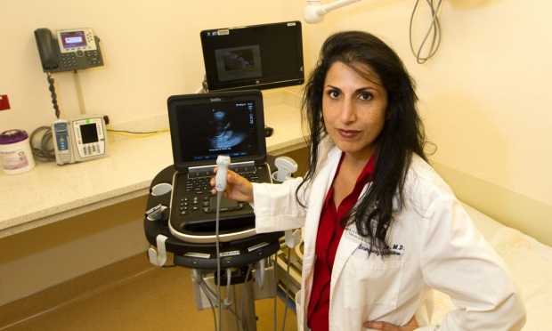
Laleh Gharahbaghian, who directs the ultrasound program in the emergency department, is seeing more community physicians using ultrasound.
Norbert von der Groeben
In the public eye, ultrasound technology is probably best embodied by the big bedside machines that enable parents to catch a revelatory glimpse of their unborn babies.
Since the 1970s, however, ultrasound has become, quietly and steadily, the Swiss Army knife of health care, with an ever-expanding repertoire of functions, based on the ability of sound waves to travel through the body and bounce back when they hit something. Now the technology has been developed into a high-resolution, often pocket-sized aid for the diagnosis and treatment of many types of injuries and medical conditions.
Ultrasound’s trajectory has been mission creep of the best possible sort.
“You name the condition, and people are trying to diagnose or treat it with ultrasound,” said Pejman Ghanouni, MD, PhD, an assistant professor of radiology at the School of Medicine who employs MRI-guided, high-intensity-focused ultrasound to treat uterine fibroids. One of the main selling points of ultrasound for medical imaging and treatment is its lack of cancer-causing radiation.
Stanford has become a center of diagnostic ultrasound research, education and training. On Oct. 18, the school will host ULTRAfest, a full day of free ultrasound instruction open to any medical student in the country. Experienced clinicians from several medical specialties will serve as teachers. Last year, more than 300 medical students from the western United States participated in the event.
The School of Medicine already has incorporated ultrasound into its anatomy training for first-year students and in its patient-doctor courses for pre-clinical students. The school will soon have a complete, four-year ultrasound curriculum, which will enable students to graduate with ultrasound competency.

A laptop ultrasound scanner.
Norbert von der Groeben
The primary goal of ULTRAfest, co-chaired by Laleh Gharahbaghian, MD, clinical associate professor of emergency medicine and director of Stanford Hospital’s emergency department ultrasound program, is to teach how ultrasound can enhance knowledge of anatomy, physiology and pathology — and, more importantly, how it can improve patient care in ways that Gharahbaghian has seen grow rapidly in the 14 years since she graduated from medical school.
“We use it for everything from head to toe and skin and organs,” she said. “It’s become an essential tool at the bedside we apply to immediately rule out — or rule in — medical conditions.”
Use in emergency medicine
Especially useful in emergency care, she said, is ultrasound’s ability “to help us find out what’s going on with a patient and to treat them appropriately with greater speed and accuracy.”
She said, “We might have a patient, for instance, who is unconscious, incoherent or not speaking a language we know, and we have no idea why the heart rate is up and the blood pressure down.”
Seeing beyond the barrier of consciousness or language is another ultrasound capability, Gharahbaghian said. “Even if a patient’s eyes are swollen shut, you can use ultrasound to quickly detect injury — to see a ruptured eye orbit or to gauge function, like pupillary activity.”
Gharahbaghian first saw this capability when the Stanford Emergency Medicine Program for Emergency Response team took along laptop-based ultrasound devices to care for people injured in the 2010 earthquake that struck Haiti. In the hardest hit areas, the earthquake destroyed most of the standing medical facilities. “In that disaster, when there was no power, when hospitals were completely collapsed and resources were limited, the ultrasound was the only radiological device the team had,” she said.
Last year, Stanford’s emergency department became the treatment center for 55 of the 200 people injured in the July 2013 crash at San Francisco International Airport of an Asiana Airlines Boeing 777. Many passengers spoke little English, and the nature of their injuries was not always immediately apparent. Seatbelts did save lives, but the violent side-to-side movement of the aircraft produced spine and rib fractures, often accompanied by seatbelt-related internal injuries caused by the unusual combination of movements in the crash. Several passengers, initially thought be only mildly injured when examined at the scene, were found, during their initial assessment with ultrasound at Stanford, to have serious internal injuries that needed quick attention.
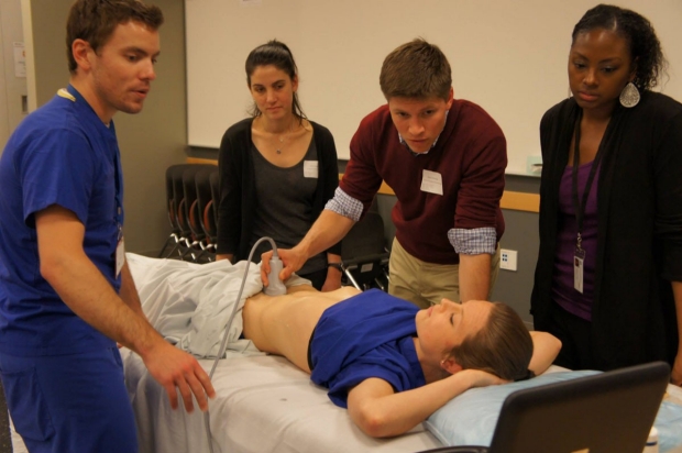
Students and teachers at last year’s ULTRAfest.
Teresa Roman-Micek
‘I just fell in love with the technology’
Almost five years ago, a few weeks before William White started his first year of medical school at Stanford, he took a class in ultrasound. “I just fell in love with the technology,” he said, “picking up a probe and looking into the body in real time.” For the next three years, still enamored with ultrasound, White continued as a volunteer assistant for the class. Now he hopes to do a residency in emergency medicine. He is also one of the organizers of ULTRAfest, an event he believes will help ultrasound overcome the unfamiliarity many medical students have with its full range of capabilities. “This current generation is starting to get very familiar with it,” he said. “I think in the future there will be a broader acceptance — and it will be part of the standard for primary care.”
That may still take some time: Fellowships that focus on ultrasound use are now only available through emergency medicine training programs, found mostly at academic medical centers like Stanford Medicine, where both basic science and clinical research is part of ultrasound’s reimagining. It also helps to have an abundance of devices: Lucile Packard Children’s Hospital Stanford has 72, still used for prenatal evaluations, but also a key part of physicians’ ability to see and treat complex cardiovascular issues.
Stanford Hospital’s 262 ultrasound devices serve an important role in emergency care, surgical treatments and postoperative care in the ICU. Ultrasound is also essential in other areas, including reproductive endocrinology and infertility, respiratory therapy, orthopaedics, anesthesia, urology, outpatient surgery, mammography, endoscopy, head and neck surgery, diagnostic radiology and cardiovascular care. Stanford’s emergency department has eight laptop-based and three handheld ultrasound devices. Ultrasound is also standard equipment at the Stanford Cancer Center, the Cath-Angio Lab and several nursing units. The Life Flight helicopter also carries ultrasound devices.
The long list of other invasive procedures made less complicated by ultrasound includes endotracheal intubation, fine needle aspiration, interventional radiology procedures, pedicle screw insertion in scoliosis surgery, prostate cancer biopsies and emergency procedures like central venous access. Ultrasound also is a much gentler and quicker screening tool for spotting artery-narrowing plaque than is coronary angiography, and for pre-operative looks at arterial issues before neck surgery.
Less costly
Ultrasound also is relatively inexpensive: Even a refurbished CT scanner with a minimum view capacity is priced at $65,000. New ones start at $90,000. Handheld ultrasounds can cost as little as $7,000; laptop-based devices range from $25,000 to $40,000.
More recently, the use of ultrasound has crossed into another part of the anatomy long thought to be immune to its imaging prowess: the lungs. In the air-filled environment of the lungs, the sound waves that are the basis of ultrasound have nothing to ping against. However, in lungs where disease has produced fluids, ultrasound has proven more accurate than a chest X-ray and faster than CT scan to diagnose common lung conditions, including pulmonary edema, pneumonia and pleural effusions.
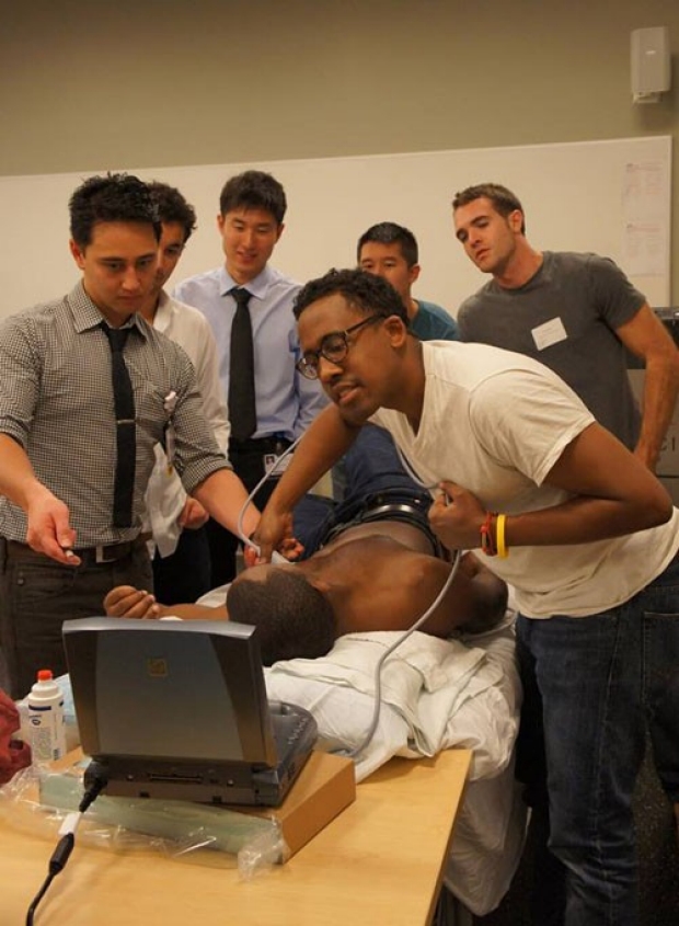
This year's full, free day of ultrasound instruction is set for Oct. 18.
Teresa Roman-Micek
Ultrasound devices at Stanford are so highly desired that “it’s not easy to keep spares,” said Harvey Fortune, assistant director of Stanford Health Care’s clinical technology group.
Ghanouni and other Stanford physician-scientists are pushing medical ultrasound to the next level. He and his colleagues, Jaimie Henderson, MD, professor of neurosurgery, and Casey Halpern, MD, assistant professor of neurosurgery, are using high-intensity-focused ultrasound, guided by MRI, to treat essential tremor, a nervous system disorder marked by uncontrollable shaking. The ultrasound heats and destroys specific brain tissue: No anesthesia, no scalp incisions, no burr holes through the skull. Another team of Stanford physicians, which includes radiologists, neurosurgeons, oncologists and physicists, plans soon to conduct an investigatory test of this technology, available only at a handful of medical centers worldwide, to allow drugs to cross the blood-brain barrier for more targeted treatment of brain tumors.
Ultrasound is an essential part of the work of Adam de la Zerda, PhD, an assistant professor of structural biology. In collaboration with Sam Gambhir, MD, PhD, professor and chair of radiology, de la Zerda recently developed and patented a technology called photoacoustic imaging that transforms light waves into ultrasound waves. Its goal is to detect cancer with a resolution that matches CT scanning and MRI.
Quick answers for patients
Viveta Lobo, MD, who completed a fellowship in ultrasound in Stanford’s emergency department and served as an ULTRAfest co-chair, said studies have shown patients feel that doctors using bedside ultrasound spend more time at patients’ bedsides.
Studies also have shown that hospitalized patients who underwent ultrasound scanning were discharged more quickly.
Ultrasound can also provide quick answers, which patients appreciate. “I can tell someone right away that there are no gallstones, or that a woman’s baby is OK,” Lobo said. Even more crucial, she said, “we can see and treat quickly that life-threatening ectopic pregnancy or large pulmonary embolism.”
But ultrasound takes some training to master, Lobo added. “You have to know how to get good images — how to move it around obstacles, like the ribs, to see what you need to see. You have to know how to adjust the settings, just as you do in photography. Then you have to know how to interpret what you’ve seen.”
Gharahbaghian is seeing more and more community physicians, as well as those in outpatient clinics, who are using ultrasound. She hopes that trend continues. “The more we spread the news of how ultrasound helps patients in all clinical settings, the better,” she said.
-
 Sara WykesSara Wykes is a writer for the Stanford Hospital & Clinics communications office. Email her at swykes@stanfordhealthcare.org.
About Stanford Medicine
Stanford Medicine is an integrated academic health system comprising the Stanford School of Medicine and adult and pediatric health care delivery systems. Together, they harness the full potential of biomedicine through collaborative research, education and clinical care for patients. For more information, please visit med.stanford.edu.

