April 7, 2014 - By Tracie White
Eight of the 10 Rodin hand sculptures on display in a new exhibit have been diagnosed for malformations and diseases by a School of Medicine hand surgeon.
One of the sculptures has been "repaired" using virtual surgery by the techies in the school's Division of Clinical Anatomy. And with the help of more digital wizardry, viewers can see virtual blood and bone in the bronze hands.
Inside Rodin's Hands: Art, Technology and Surgery, which runs April 9 through Aug. 3 at Stanford's Cantor Arts Center, is a feat of interdisciplinary collaboration that celebrates the connection between sculptor Auguste Rodin's fascination with the human form and medicine's fascination with human anatomy.
"A deep and rich history unites the art of the museum with the medical school," said Connie Wolf, director of the Cantor Arts Center, which has one of the largest Rodin collections, with 200 of his sculptures including The Thinker and The Gates of Hell. "These statues have inspired faculty at the School of Medicine. Art is informing medicine in the exhibit, and medicine is informing art. It's unlike anything we've ever done before."
Situated near the medical school and Stanford Hospital, the Rodin collection at the Cantor Arts Center has for decades been a magnet for surgeons, physicians and other students of anatomy. One, hand surgeon James Chang, MD, professor and chief of plastic and reconstructive surgery, has regularly strolled from hospital to museum since his days as a surgery resident 15 years ago. His long fascination with Rodin's hand sculptures is the inspiration for this new exhibit, Wolf said.
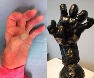
The hand of a patient with Charcot-Marie-Tooth disease6, an inherited neurological disorder, resembles Rodin's Large Clenched Left Hand. Both images appear in an iBook developed to accompany the exhibit Inside Rodin's Hands, on view April 9 through Aug. 3 at the Cantor Arts Center.
"As a surgery resident, I began to notice that most of the hands looked like the conditions I was treating, from fractures to malformations to tumorous growths," Chang said.
As something of a game or a hobby, he began to catalog these specific conditions. He thought the sculpture Large Left Hand appeared to have some broken metacarpals. Rodin's Large Clenched Hand, a sculpture frozen in a painfully exaggerated and abnormal posture, had Charcot-Marie-Tooth disease, an inherited neurological disorder, he speculated. From sculpture to sculpture, hand to hand, the surgeon has identified a ganglion cyst, a thumb amputation, a stiff joint and other conditions.
Chang's personal hobby — complete supposition — eventually turned into a teaching tool, which he incorporated into the undergraduate course Surgical Anatomy of the Hand: From Rodin to Reconstruction. Now he regularly brings his students, and his surgical trainees, to the museum to study anatomy using the sculptures.
Evaluations by Chang and his students of the bronze hands' perceived conditions will accompany the exhibit. The Large Left Hand sculpture that seemingly has a few fractured metacarpals has been virtually operated on. By combining 3-D printing with engineering skill, a team from the Division of Clinical Anatomy produced a model of the sculpture as it would look after surgical success.
The team used actual patient CT scans from the hand clinic to create 3-D models of the supposed internal anatomies of Rodin's hands and placed them into the virtual sculptures. Visitors can see the anatomy that lies beneath the "skin" of the bronze hands by looking at the sculptures through an iPad.
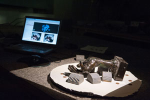
Three-dimensional scanning technology was used to digitally map the external surface of Rodin's hand sculptures.
During a recent visit to the museum, Chang pointed to a hand on the sculpture titled The Three Shades. "See the fist? The skin begins to fuse together. 'Epidermolysis bullosa,' a connective tissue disease, is the first thing I thought of. This is what draws me to these sculptures."
No one knows for certain whether Rodin's hand sculptures were based on living models or grew from his imagination. He certainly studied anatomy like other art students of his day, and it is known that he spent time at the Musée Dupuytren in Paris (a museum of anatomical items illustrating diseases and malformations), according to Bernard Barryte, a Rodin expert and the curator at the Cantor Center who worked on the new exhibit.
"Apparently, Rodin hung out not only with writers, but also with several scientists," Barryte said. "He was all about nature. He tried not to use conventional poses. He was not trying to create the perfect hand, but the truly human hand."
The relationship between art and anatomy does have a rich history, stretching back to Renaissance artists such as Michelangelo and Leonardo da Vinci, who conducted their own anatomical dissections to inform their art — contributing to science along the way.
Stanford has its own history of scientists who have used art to inform their teaching and learning, stretching back to Robert Chase, MD, the Emile Holman Professor of Surgery, Emeritus, and founder of the Robert A. Chase Hand & Upper Limb Center.

A rendering of Rodin's Left Hand of Eustache de St. Pierre, in which you can see virtual blood vessels and bone with the help of an iPad at the exhibit.
"I think the modern-day vision of bringing to life art and anatomy is in the form of Bob Chase," said Amy Ladd, MD, professor of orthopaedic surgery and chief of the hand and upper limb center, in a video about the exhibit. "He was kind of the ring leader in making sure anatomists and artists talk to each other."
Part of the new Rodin exhibit builds upon this unique history, according to Paul Brown, DDS, consulting associate professor of anatomy, who together with exhibit production manager Matt Hasel and medical artist Sarah Hegmann have used technology to allow visitors to see inside Rodin's hands.
"We would visit the museum with Bob Chase, and he would bring the statues to life," Brown said. "We had discussed at length the idea of doing a three-dimensional representation of them, scanning them in somehow.
"Two years ago, as a summer project, we did just that — created virtual replicas of the hands." This exhibit is an extension of that early work.
"Anatomy really comes alive in 3-D," Chang said. "I tell my residents to always remember that hand surgery is three-dimensional."
That's why the Rodin sculptures are so helpful in teaching anatomy to his students and his surgical trainees, Chang said. And, now, to the public as well. "With this innovative exhibit we hope all will see and enjoy the Rodin hands in an entirely new way," he said.
-
 Tracie WhiteTracie White is a science writer for the medical school’s Office of Communication & Public Affairs.
About Stanford Medicine
Stanford Medicine is an integrated academic health system comprising the Stanford School of Medicine and adult and pediatric health care delivery systems. Together, they harness the full potential of biomedicine through collaborative research, education and clinical care for patients. For more information, please visit med.stanford.edu.

