February 10, 2014 - By Amy Adams
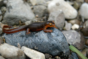
In the 1960s, Stanford scientists discovered that a toxic chemical in the skin and eggs of California newts was identical to toxin in pufferfish. Now, scientists are using a modified version of a similar toxin to pinpoint the source of pain in laboratory rats.
The California newts on the Stanford campus may be limited in physical range to the area around a small, now-dry lake, but their sphere of scientific influence extends much further. Work that started in these amphibians decades ago has now resulted in a tool that for the first time can highlight the location of pain generation in a living animal.
The work began in the 1960s, when Stanford scientists discovered that the native newts at Stanford had a chemical in their skin and eggs identical to the potent toxin found in pufferfish. (Outdoorsy students and their parents need not be afraid; the newts are only toxic if eaten.)
The late Harry Mosher, PhD, a professor of chemistry, spent much of his career analyzing and attempting to synthesize this toxin and related molecules, collectively called guanidinium toxins. They turned out to be an interesting group of molecules for chemists who like to develop new ways of stitching atoms together to form molecules. One such chemist, Justin Du Bois, PhD, later joined Stanford and started a lab focused on making and modifying the structures of the guanidinium toxins, even inheriting some chemicals from Mosher's lab.
Du Bois, an associate professor of chemistry, didn't initially know about Mosher's early work, but when he did they had a conversation that sent Du Bois in a new direction. In nature, pufferfish, frogs and many other animals are poisonous because the toxins they contain bind to channels on the surface of nerves and prevent those nerves from firing.
Blocking pain neurons
Mosher pointed out that if someone could create a version of the toxin that only latched onto the channels in nerves that signal pain, the chemical could conceivably block pain neurons — therefore blocking the sensation of pain — without interfering with other nerves like the ones that signal muscles to move.
"He was the one who first said to me that if there was some way of localizing the site of action of the drug it would make for a great pain medicine," Du Bois said. "I thought if we can make and modify it and make it stay at the site of the injection, we would really have something."
Du Bois started reading up on nerve physiology and talking to biologists about pain. Meanwhile, across the street from where Du Bois and his students were getting ever better at synthesizing guanidinium toxin variants, a new radiologist on campus, Sandip Biswal, MD, was growing frustrated with his inability to diagnose the cause of pain.
Biswal, an associate professor of radiology at the School of Medicine, spent a lot of time imaging parts of the body where people said they felt pain, trying to find the source. It was a frustrating task because often the source of pain isn't obvious, and sometimes the source is far removed from where a person feels the sensation of pain. Other times, he'd see something that looked painful, surgeons would fix it, and the patient would still be in pain.
"All this made me think that we needed to be able to image pain more accurately," Biswal said. "It would be great if we could take a picture of something that is actively sending pain signals up to the brain."
Biswal and Du Bois met, realized their overlapping interests and obtained funding from the Bio-X program to work together. (Bio-X supports this kind of interdisciplinary collaboration.) Their initial goal was to modify guanidinium toxins to bind neurons signaling pain to give off a signal visible outside the body. (They recently published the findings of this work in the Journal of the American Chemical Society.)
Du Bois worked with Frederick Chin, PhD, an assistant professor of radiology, to create a modified version of guanidinium toxins that contained a non-toxic molecular tag commonly used for imaging chemicals in the body. With this molecular flag, scientists would be able to trace the location of this modified toxin in an animal.
Success in rats
The group tested the new compound in rats that had leg injuries. After injecting the compound into these animals, they could see it located in the leg that had a nerve injury but not in the opposite, uninjured leg. The chemical had latched onto those nerves signaling pain.
Du Bois said that although the results show potential for human applications, much needs to be done to translate work from rodents to humans. The group has formed a company to refine the compound so it binds even more specifically to neurons conducting pain and to ensure that it will be safe to test in people. The group members hope the work will one day help doctors locate the source of a patient's pain and also perhaps produce a new class of drug for treating pain.
In addition to being an example of what can happen when scientists from across campus work together, Du Bois and Biswal say the research specifically shows the role molecular scientists can play in solving biological problems. Du Bois is on the executive committee for the new Stanford Institute for Chemical Biology, which was formed specifically to encourage collaborations like this one.
"The beauty of Stanford is that you can do projects like this," Du Bois said. "If I couldn't walk across the street and engage with my friends like Sandip in real time, I am certain our program in pain research would have never taken off."
-
 Amy AdamsAmy Adams is the director of interdisciplinary life sciences communications for Stanford University.
About Stanford Medicine
Stanford Medicine is an integrated academic health system comprising the Stanford School of Medicine and adult and pediatric health care delivery systems. Together, they harness the full potential of biomedicine through collaborative research, education and clinical care for patients. For more information, please visit med.stanford.edu.

