March 21, 2013 - By Krista Conger

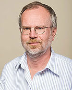
Roeland Nusse
Cells in the body need to be acutely aware of their surroundings. A signal from one direction may cause a cell to react in a very different way than if it had come from another direction. Unfortunately for researchers, such vital directional cues are lost when cells are removed from their natural environment to grow in an artificial broth of nutrients and growth factors.
Now, researchers at the Stanford University School of Medicine and the Howard Hughes Medical Institute have devised a way to mimic in the laboratory the spatially oriented signaling that cells normally experience.
Using the technique, they’ve found that the location of a “divide now” signal on the membrane of a mouse embryonic stem cell governs where in that cell the plane of division occurs. It also determines which of two daughter cells remains a stem cell and which will become a progenitor cell to replace or repair damaged tissue.
The research offers an unprecedented, real-time glimpse into the intimate world of a single stem cell as it decides when and how to divide, and what its daughter cells should become. But the implications stretch beyond stem cells.
“In the body, it is likely that every cell grows and differentiates in some kind of orientation,” said Roeland Nusse, PhD, professor of developmental biology. “Without this guidance, specialized cells would end up in the wrong place. Now, we can study the division of single mammalian cells in real time and see them dividing and differentiating in an oriented way.
Understanding this process of self-renewal and specialization (or differentiation) is critical to learning how to truly harness the power of stem cells for future therapies. But polarity, or the ability of a cell to distinguish its top from bottom or left from right, is also vital to many other biological processes. For example, hair grows out of, rather than into, the body, and tissues develop with orderly layers of specific cell types.
Nusse is the senior author of the work, published March 23 in Science. He is also a member of the Stanford Cancer Institute, the Stanford Institute for Stem Cell Biology and Regenerative Medicine and HHMI investigator. Shukry Habib, PhD, a research associate and Siebel Scholar, is the lead author of the work. The study was funded in part by a grant from the California Institute of Regenerative Medicine.
Stem cells are unique in their ability to both self-renew and to generate progenitor cells that can become many cell types. A single stem cell can divide to make two new stem cells or, in a process called asymmetric division, give rise to one stem cell and one progenitor cell. Because the original parent cell is replaced by the two new daughter cells, this approach ensures that stem cells will not be depleted during periods of development or healing.
Things change when these cells are grown in the laboratory, however. Researchers usually choose growth conditions that favor specific outcomes: self-renewal or differentiation into specialized cell types. These growth or differentiation signals affect all parts of the cell equally, and it’s not been possible under these conditions to ascertain whether and how the location of these signals may affect the outcome.
In the current study, Habib tested the effect on mouse embryonic stem cells of a protein called Wnt3a, which is known to play a critical role in embryonic development and in the growth and maintenance of stem cells. The stem cells have many receptors for Wnt proteins on their surfaces, and Wnt3a has been shown to promote self-renewal over differentiation in several types of stem cells.
Habib attached molecules of Wnt3a protein to tiny synthetic beads and incubated the beads with embryonic stem cells. He then observed the reaction over time of the cells to which a single bead-bound protein had attached via one of the cell’s many Wnt3a receptors.
The effect of the localized signal was clear. In 75 percent of cases, the stem cell began to divide in a very specific orientation, with the plane of division occurring perpendicularly to the location of the incoming signal. In contrast, only 12 percent of cells exposed to beads bound to a control protein exhibited similar patterns of division.
Habib and his colleagues also found that the daughter cell closest to the Wnt3a signal expressed proteins showing it was maintaining its pluripotency, or ability to function as a stem cell like its parent. The one farthest from the signal, however, expressed proteins indicating that it was beginning to differentiate.
The researchers speculate that the reduction in the intensity of the Wnt signal in the distant daughter cell is what causes it to begin the differentiation process; the loss of the Wnt3a signal is known to cause cultured stem cells to begin differentiating.
“We found that these two processes, division and differentiation, are coupled,” said Nusse, who is also the Virginia and Daniel K. Ludwig Professor in Cancer Research. “In real life, of course, the cell is exposed to many signals simultaneously. But by studying just one protein and just one cell, we can clearly see that the cell’s division is aligned to the source of the Wnt signal. One of the daughters is always closer and remains pluripotent. The other is further from that signal; it begins to differentiate.”
The researchers would like to extend their studies to other types of stem cells, as well as to cells that have mutations in other components known to affect cell polarity.
Other Stanford co-authors include postdoctoral scholar Feng-Chiao Tsai, MD, PhD, and professor of chemical and systems biology Tobias Meyer, PhD.
The research was funded by the National Institutes of Health, CIRM, HHMI and the Center for Regenerative Therapies in Dresden. Habib and Nusse have applied for a patent on the bead immobilization technique described in the paper, as well as its applications.
Information about the Department of Developmental Biology, which also supported the work, is available at http://devbio.stanford.edu.
About Stanford Medicine
Stanford Medicine is an integrated academic health system comprising the Stanford School of Medicine and adult and pediatric health care delivery systems. Together, they harness the full potential of biomedicine through collaborative research, education and clinical care for patients. For more information, please visit med.stanford.edu.